Neuroplasticity & Neurotrophins
How the brain physically rewires itself — the glutamate synapse, the molecular logic of "fire together, wire together", and the family of growth proteins (BDNF the star among them) that decides which connections grow and which fade. The mechanistic foundation under every "plasticity stack", every nootropic peptide, and the honest limits of what they can do.
A short history first, because the central idea was disbelieved for most of a century.
In the 1890s the Spanish anatomist Santiago Ramón y Cajal established the neuron doctrine — the brain is built from discrete cells (neurons) that communicate across gaps, not a continuous mesh. It won him a Nobel Prize and founded modern neuroscience. But Cajal also drew the wrong conclusion about change. In 1928 he wrote that in the adult brain "the nerve paths are something fixed, ended, immutable. Everything may die, nothing may be regenerated." For fifty years that was orthodoxy: you were born with your neurons and your wiring, and the only direction was decline.
The cracks came slowly. In 1949 the psychologist Donald Hebb proposed, in The Organization of Behavior, that learning is physical — "when an axon of cell A is near enough to excite cell B and repeatedly takes part in firing it, some growth process takes place such that A's efficiency in firing B is increased." This is Hebbian plasticity, compressed into the slogan "cells that fire together, wire together." It was a hypothesis with no known mechanism. The mechanism arrived in 1973, when Tim Bliss and Terje Lømo stimulated the rabbit hippocampus and found that a brief burst of high-frequency activity made a synapse stronger for hours — long-term potentiation (LTP), the first concrete demonstration that a synapse remembers. Then in 1982 Yves-Alain Barde and Hans Thoenen purified, from pig brain, a tiny protein that kept neurons alive and growing: brain-derived neurotrophic factor (BDNF) — the molecule that turns out to sit at the centre of how experience, exercise, and many of the compounds in this database act on the brain.
The modern picture is the opposite of Cajal's pessimism: the adult brain remodels itself continuously, at every level from the single synapse to (controversially) the birth of entirely new neurons. The frontier today is twofold — neurotrophin-mimetic drugs that try to bottle BDNF's effect in a small, brain-penetrant molecule (the entire premise of Cerebrolysin, Semax, dihexa, and eutropoflavin), and a still-unsettled argument over whether the adult human brain makes new neurons at all. This page builds the whole system from first principles so that both frontiers make sense.
Why this page exists
The biohacking world is awash in the word plasticity. People take Semax "for BDNF", Lion's Mane "for NGF", dihexa "for synaptogenesis", and intranasal insulin "for neurogenesis", mostly without a model of what any of those words mean or how the steps connect. The result is that obviously different mechanisms — making a single synapse temporarily stronger, growing a new dendritic spine, transcribing a survival gene, birthing a new cell in the hippocampus — get blurred into one fuzzy promise of "rewiring your brain".
This page draws the real map. We start with what plasticity is and its distinct levels, then build the glutamatergic synapse from scratch, derive LTP and LTD (the cellular events behind learning) from the behaviour of two receptors, then move up to structural remodelling of the neuron, then to the neurotrophins — the growth proteins, BDNF chief among them, that gate the whole process — and finally to adult neurogenesis, where we calibrate honestly against a genuinely contested literature. By the end you should be able to take any "plasticity" compound and say which level it acts on and through which molecule — and therefore what it can and cannot plausibly do.
This is the sibling of the Neuroscience of Cognition page, and the two divide the territory cleanly. That page owns dopamine — its synthesis, its three pathways, the reuptake transporters, the stimulants, and the prefrontal inverted-U. This page owns plasticity and the neurotrophins — glutamate, the NMDA receptor, LTP, BDNF, and neurogenesis. Where they meet (the synapse as a concept, the NMDA receptor's magnesium block) we cross-link rather than repeat.
The four levels of plasticity
"Neuroplasticity" is not one phenomenon. It is a stack of four, operating on different timescales and through different machinery. Keeping them separate is the single most clarifying move in the whole subject.
- Synaptic plasticity — changing the strength of an existing connection between two neurons, by making the receiving side more or less responsive. This is the fastest level (seconds to hours to set up) and the cellular basis of learning and memory. LTP and LTD live here.
- Structural plasticity — physically growing or removing the connecting hardware: new dendritic spines (the tiny protrusions that receive synapses), pruning of unused ones, sprouting of new axon branches. Slower (hours to days), and it is how a strengthened synapse becomes a permanent one.
- Functional plasticity / remapping — the brain reassigning jobs across regions: after a stroke, neighbouring cortex takes over a lost function; a blind person's visual cortex gets recruited for touch and hearing. This is the large-scale, systems-level consequence of the two levels below it.
- Neurogenesis — the birth of entirely new neurons from neural stem cells. In the adult mammal this is confined to one or two niches (chiefly the hippocampus), and in adult humans its very existence is debated — we treat it last and carefully.
The key relationship runs upward: synaptic changes, if reinforced, are consolidated into structural changes, and enough structural change across a population produces functional remapping. Neurogenesis feeds raw new units into the bottom of the hippocampal version of this stack. A compound that "boosts plasticity" must be pinned to a level before the claim means anything.
The glutamatergic synapse: the unit of learning
Almost all fast excitatory signalling in the brain — and essentially all of the plasticity that underlies learning — happens at synapses that use the neurotransmitter glutamate (the anion of the amino acid glutamic acid, the brain's principal excitatory messenger). To understand LTP you must first understand this one synapse in detail.
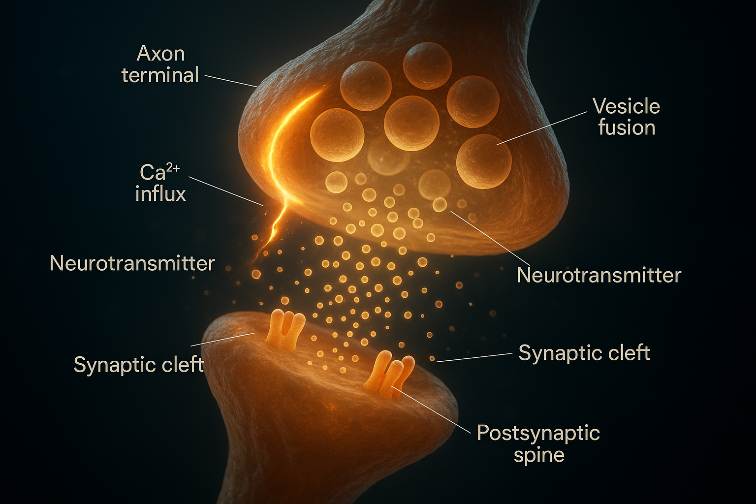 The synapse as a working system: an impulse reaches the axon terminal and opens calcium channels; the calcium triggers vesicles to fuse and dump neurotransmitter into the cleft; it crosses and opens receptor channels on the postsynaptic spine. The flat diagram further down zooms in on the two receptors — AMPA and NMDA — that make this particular synapse learn.
Recall the basic synapse from the cognition page: the upstream presynaptic neuron releases a neurotransmitter into the synaptic cleft (the gap), and it binds receptors on the downstream postsynaptic neuron. At a glutamatergic synapse the postsynaptic membrane carries two kinds of glutamate receptor sitting side by side, and the entire logic of learning falls out of the difference between them.
The AMPA receptor (named for a synthetic molecule, α-amino-3-hydroxy-5-methyl-4-isoxazolepropionic acid, that selectively activates it) is the workhorse. It is a ligand-gated ion channel: glutamate binds, the channel opens immediately, and sodium ions (Na⁺) flood in, depolarising the postsynaptic cell (making its interior less negative). This is the ordinary, millisecond-by-millisecond business of excitatory transmission. The number of AMPA receptors a synapse displays in its membrane is, to a first approximation, the strength of that synapse — more AMPA receptors means a bigger response to the same puff of glutamate. Hold that fact; it is the punchline of LTP.
The NMDA receptor (named for N-methyl-D-aspartate) is the clever one — and it is the coincidence detector at the heart of Hebbian learning. It has three features that conspire:
- Like AMPA, it binds glutamate and is an ion channel. But it has an extra requirement: it also needs a co-agonist, glycine or D-serine, to be present — a permissive "the network is ready" signal.
- Crucially, at the cell's normal resting voltage the NMDA channel is physically plugged by a magnesium ion (Mg²⁺) sitting in its pore. Glutamate can bind all it likes; the channel stays blocked. (This magnesium plug is the subject of the Magnesium deep dive — it is what makes the receptor a clean signal-to-noise filter.)
- The plug is voltage-dependent. Only when the postsynaptic membrane is already depolarised — pushed positive by strong AMPA-receptor activity nearby — is the Mg²⁺ electrostatically expelled from the pore.
- And the NMDA channel, once unblocked, is permeable not just to Na⁺ but to calcium ions (Ca²⁺) — and Ca²⁺ inside a neuron is not merely charge, it is a second messenger, a chemical command signal that switches on enzymes.
Put those together and the NMDA receptor opens its calcium gate only when two things happen at once: glutamate is present in the cleft (the presynaptic neuron fired) AND the postsynaptic membrane is already depolarised (the postsynaptic neuron is also active, expelling the Mg²⁺). That is a molecular AND-gate — a coincidence detector. It is the physical implementation of Hebb's rule: the synapse strengthens precisely when pre- and postsynaptic cells are active together. "Fire together, wire together" is not a metaphor; it is the magnesium block.
flowchart TD
GLU["Glutamate in cleft<br/>(presynaptic fired)"] --> AND{NMDA AND-gate}
DEP["Postsynaptic depolarised<br/>(AMPA-driven, cell active)"] --> AND
DEP -->|"expels Mg2+ plug"| AND
AND -->|"both true at once"| OPEN["NMDA channel opens"]
AND -->|"only one true"| SHUT["Stays blocked<br/>(no signal)"]
OPEN --> CA["Ca2+ floods in<br/>= second-messenger command"]
CA --> PLAST["Plasticity triggered"]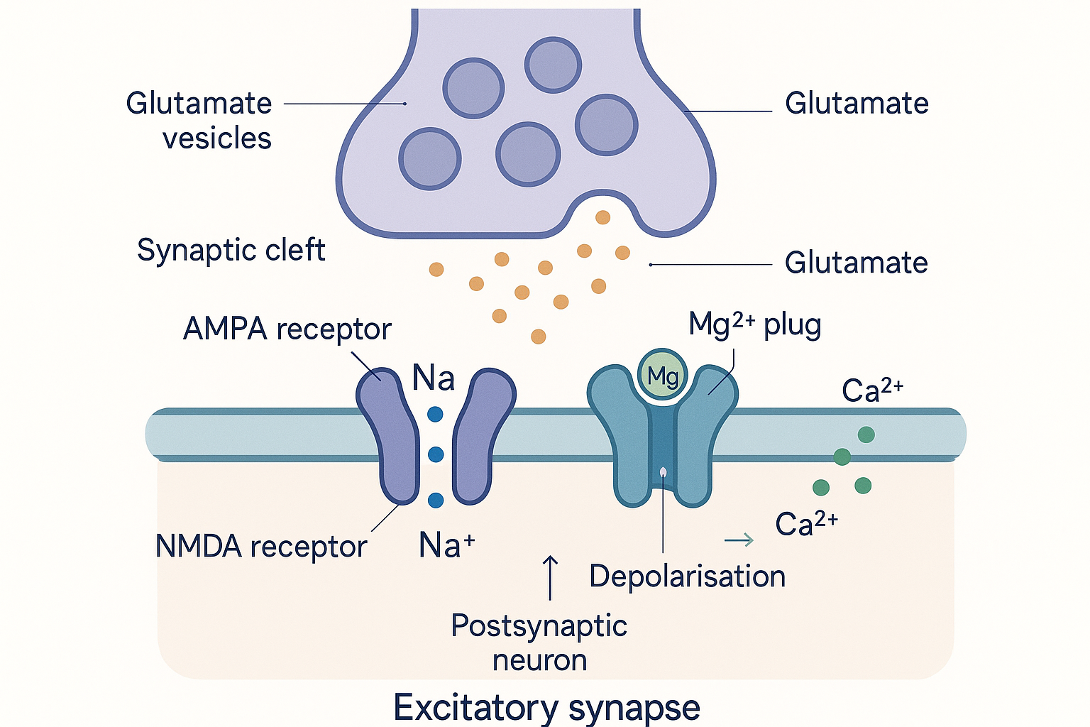 The glutamatergic synapse, the unit of learning: AMPA receptors carry the everyday sodium signal, while the NMDA receptor stays plugged by magnesium until the cell is both bound by glutamate and already depolarised — only then does its calcium gate open. That calcium is the trigger for everything downstream.
Long-term potentiation: how a synapse remembers
Long-term potentiation (LTP) is a long-lasting increase in synaptic strength following a brief burst of strong, high-frequency activity — exactly the Bliss-and-Lømo phenomenon. It is the leading cellular model of how memories are physically stored. It comes in two phases, and the distinction matters because different compounds engage different phases.
Induction is the calcium spike. A high-frequency burst drives strong AMPA-mediated depolarisation, which expels the NMDA magnesium plug, which lets a large, sharp pulse of Ca²⁺ into the postsynaptic spine. The amount and timing of calcium is the whole instruction set — a big fast spike means "strengthen" (LTP); a small slow trickle means "weaken" (LTD, below). Same ion, opposite outcomes, read off the dose.
Early-LTP (E-LTP) is the first 1–3 hours and needs no new proteins — it works by modifying machinery that is already there. The calcium pulse activates a remarkable enzyme, CaMKII (calcium/calmodulin-dependent protein kinase II), which is abundant at the synapse and has a special trick: once switched on it phosphorylates itself (autophosphorylation at threonine-286), so it stays active even after the calcium subsides. It is a molecular switch that flips and latches — a tiny memory element in protein form. Active CaMKII then does two things that directly raise synaptic strength:
- It phosphorylates existing AMPA receptors, increasing their single-channel conductance (each one passes more current).
- It drives the trafficking of additional AMPA receptors out of internal stores and into the postsynaptic membrane, from the surrounding membrane into the synapse itself.
Recall that AMPA-receptor number is synaptic strength. So E-LTP is, at its core, inserting more AMPA receptors so the same glutamate now produces a bigger response. The synapse is louder.
Late-LTP (L-LTP) is what turns a louder synapse into a lasting one. Beyond a few hours, maintaining the change requires new protein synthesis and gene transcription. The signalling cascades (especially the MAPK/ERK pathway, which we meet again under BDNF) carry the signal to the nucleus and switch on the transcription factor CREB (cAMP-response-element-binding protein) — the master "write to long-term storage" switch. CREB turns on plasticity genes (including BDNF itself, and Arc), whose protein products are captured at the activated synapse and used to physically rebuild and enlarge it — the structural step in the next section. This is why consolidating a memory takes time and is blocked by protein-synthesis inhibitors, and why sleep, exercise, and BDNF — all of which feed this transcriptional step — are the real levers on durable learning.
flowchart LR
    HFS["High-frequency burst"] --> CASPIKE["Large Ca2+ spike<br/>via NMDA receptor"]
    CASPIKE --> CAMK["CaMKII activated<br/>+ self-phosphorylates<br/>(latches ON)"]
    CAMK --> PHOS["Phosphorylate existing<br/>AMPA receptors"]
    CAMK --> INSERT["Insert NEW AMPA<br/>receptors into membrane"]
    PHOS --> ELTP["Early-LTP<br/>synapse is louder<br/>(no new protein, 1-3h)"]
    INSERT --> ELTP
    ELTP --> ERK["ERK/MAPK to nucleus"]
    ERK --> CREB["CREB switches on<br/>plasticity genes (Arc, BDNF)"]
    CREB --> LLTP["Late-LTP<br/>new protein, structural<br/>rebuild, lasts days+"]
Long-term depression and the need to forget
A brain that could only strengthen would saturate — every synapse would climb to maximum and the network would lose all contrast, like a photograph over-exposed to pure white. Learning requires the opposite operation too. Long-term depression (LTD) is a long-lasting weakening of a synapse, and it is just as important: it prunes noise, corrects errors, and frees capacity.
The elegant part is that LTD uses the same NMDA receptor and the same calcium signal as LTP — it simply reads a different pattern of it. Where LTP is driven by a large, brief calcium spike (from high-frequency input), LTD is driven by a modest, prolonged calcium rise (from weak, low-frequency input). That smaller, slower calcium signal preferentially activates protein phosphatases — enzymes that remove phosphate groups, the chemical opposite of the kinases in LTP — chiefly calcineurin (PP2B) and PP1. These dephosphorylate AMPA receptors and trigger their removal (internalisation) from the membrane. Fewer AMPA receptors means a weaker synapse. LTP inserts AMPA receptors; LTD removes them. The synapse's strength is, at bottom, a dynamic count of how many AMPA receptors it is currently displaying, set by the tug-of-war between calcium-activated kinases and phosphatases.
This is also where the homeostatic problem appears. Pure Hebbian rules are unstable — strong synapses get stronger and grab more activity, which strengthens them further, a runaway loop. The brain counters this with homeostatic plasticity, two mechanisms worth naming:
- Synaptic scaling (Gina Turrigiano's work): a neuron monitors its own average firing rate and, if it drifts too high or low over hours, scales all of its synapses up or down multiplicatively to bring itself back to target — preserving the relative pattern (the learned information) while resetting the overall gain. It is automatic gain control.
- Metaplasticity ("the plasticity of plasticity"): prior activity shifts the threshold at which future LTP versus LTD is induced. A synapse that has been very active recently becomes harder to potentiate and easier to depress, and vice versa — a sliding set-point (the BCM theory) that keeps the system in its sensitive operating range.
Together these explain why you cannot simply "max out" plasticity with a compound: the brain actively defends a set-point, and crude, network-wide BDNF or glutamate pushes invite the homeostatic machinery (and, at the extreme, excitotoxicity — calcium overload killing the cell) to push back. Useful plasticity is targeted calcium at the right synapses, not a flood everywhere.
Structural plasticity: rebuilding the hardware
A strengthened synapse is, at first, just a chemical state — more AMPA receptors, latched CaMKII. To persist, that change is written into the physical shape of the neuron. This is structural plasticity, and it centres on the dendritic spine.
A dendrite is one of the branching "input" cables of a neuron. Its surface is studded with thousands of dendritic spines — tiny mushroom-shaped protrusions, each typically hosting a single excitatory synapse on its bulbous head. The spine is the physical unit of a memory, and its size tracks its strength: spines come as small thin "learning-ready" spines and large mushroom "stable, strong" spines. The currency converting one to the other is the actin cytoskeleton — actin is the protein that polymerises into filaments forming a cell's internal scaffolding, and a spine is essentially a knot of dynamic actin under a membrane.
When LTP fires, the calcium/CaMKII signal also drives actin polymerisation inside the spine, and the spine physically enlarges — its head swells, its surface area grows, and that larger head accommodates and anchors the newly inserted AMPA receptors. A thin spine matures into a mushroom spine. This structural enlargement, stabilised by the new proteins from the L-LTP transcription step, is what converts a transient potentiation into a durable one. Conversely, LTD shrinks spines, and chronically unused spines are pruned away entirely — eliminated, their synapse dissolved. (Large-scale developmental spine pruning in adolescence, refining the cortex, is the same machinery turned up.)
Beyond the spine, structural plasticity includes axonal sprouting — the output fibre of a neuron growing new branches and boutons to form connections with new partners — which is slower and more limited in the adult brain but is central to recovery after injury and to the functional remapping mentioned earlier. The neurotrophins are the chemical signals that gate all of this growth: actin remodelling, spine stabilisation, and axonal sprouting are downstream of exactly the BDNF/TrkB signalling we turn to now.
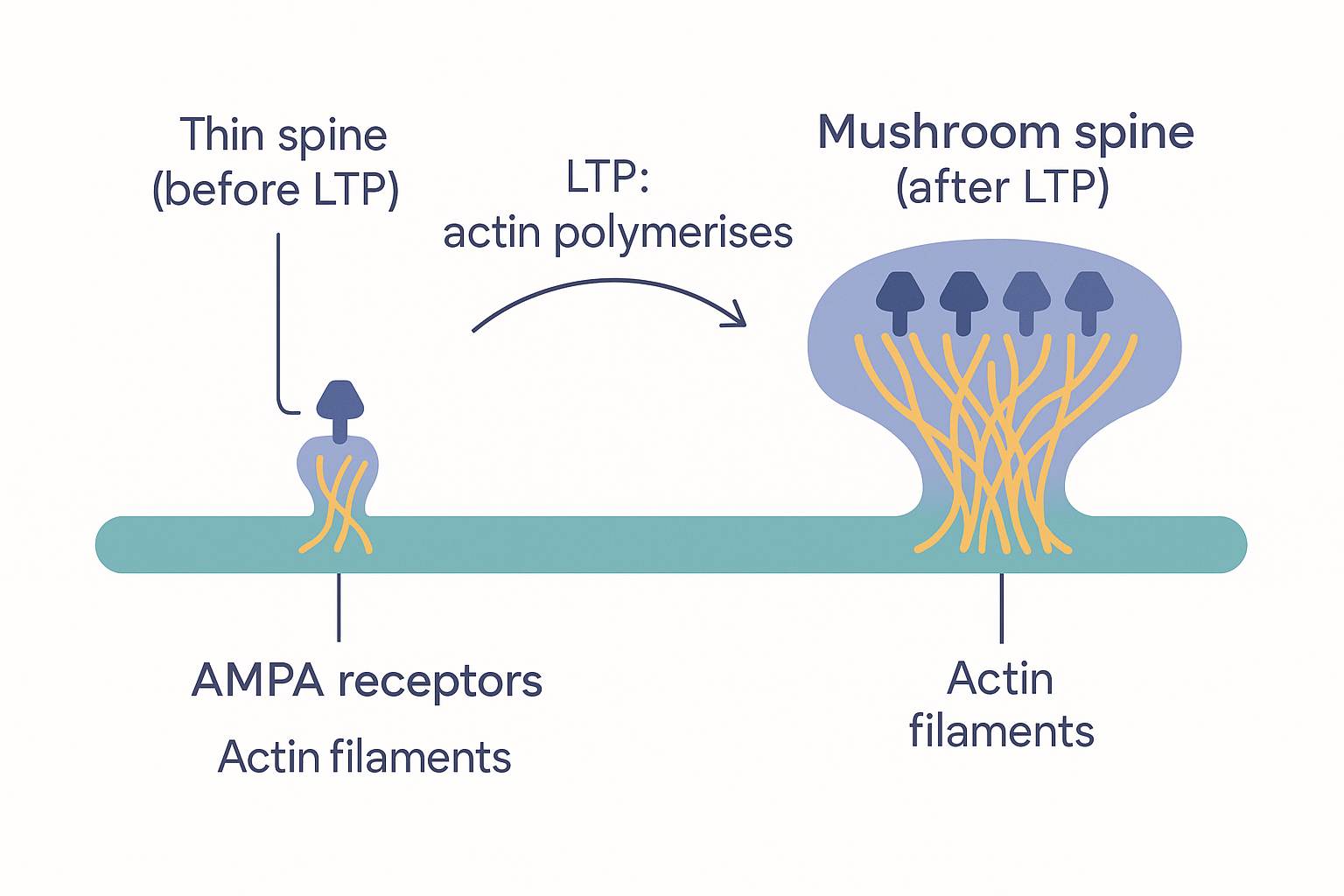 Structural plasticity makes a memory physical: LTP drives actin to polymerise inside the dendritic spine, swelling a small thin spine into a large mushroom spine whose enlarged head anchors the newly inserted AMPA receptors. Spine size is, quite literally, synaptic strength made visible.
The neurotrophins: the brain's growth and survival proteins
We have reached the proteins that decide which neurons live, which synapses grow, and which connections are reinforced. The neurotrophins are a small family of secreted growth factors — signalling proteins that bind receptors on a target cell and instruct it to survive, grow, or change. There are four in mammals, discovered across four decades:
- NGF (nerve growth factor) — the founding member, discovered by Rita Levi-Montalcini and Stanley Cohen in the 1950s (Nobel Prize 1986). Supports sensory and sympathetic neurons and the basal-forebrain cholinergic neurons that degenerate in Alzheimer's. (NGF is the headline mechanism behind Lion's Mane mushroom claims.)
- BDNF (brain-derived neurotrophic factor) — Barde's 1982 protein, and the star of this page. The dominant neurotrophin in the cortex and hippocampus, the principal regulator of LTP, learning, and mood-related plasticity.
- NT-3 (neurotrophin-3) and NT-4 (neurotrophin-4, also called NT-4/5) — the two later, less-studied members, important in development and in specific neuron populations.
Each is made as a larger precursor, a pro-neurotrophin (e.g. proBDNF), which is cleaved to the mature form. This is not a trivial detail: the pro- and mature forms can have opposite effects (a "yin-yang"), and they act through different receptors.
The receptors are the key to specificity. There are two receptor systems:
- The Trk receptors (tropomyosin receptor kinases, "track") — TrkA, TrkB, TrkC — are the high-affinity receptors that carry the positive, growth-and-survival signal of the mature neurotrophins. They are receptor tyrosine kinases: the receptor's own intracellular tail is an enzyme. Each Trk has its preferred ligand:
| Neurotrophin | Primary Trk receptor | Headline role |
|---|---|---|
| NGF | TrkA | sensory/sympathetic & basal forebrain cholinergic neurons |
| BDNF, NT-4 | TrkB | cortical/hippocampal plasticity, LTP, mood |
| NT-3 | TrkC (also TrkA/B weakly) | development, proprioceptive neurons |
- The p75 receptor (p75^NTR^) binds all neurotrophins with low affinity, and binds the pro-forms best. On its own it tends to signal the opposite: it can drive apoptosis (programmed cell death), promote LTD, and prune connections. So the same family contains both the "grow and strengthen" signal (mature neurotrophin → Trk) and the "retract and die" signal (pro-neurotrophin → p75). Which one wins depends on which form is released and which receptor dominates — a built-in sculpting system that both builds and removes.
flowchart TD
    subgraph MATURE["Mature neurotrophins to Trk = GROW / SURVIVE / STRENGTHEN"]
        NGF[NGF] --> TRKA[TrkA]
        BDNF[BDNF] --> TRKB[TrkB]
        NT4[NT-4] --> TRKB
        NT3[NT-3] --> TRKC[TrkC]
    end
    subgraph PRO["Pro-neurotrophins to p75 = RETRACT / PRUNE / APOPTOSIS"]
        PROB["proBDNF, proNGF"] --> P75["p75 receptor"]
    end
    TRKA --> GROW["Survival, growth,<br/>LTP, spine enlargement"]
    TRKB --> GROW
    TRKC --> GROW
    P75 --> DIE["Apoptosis, LTD,<br/>spine pruning"]
BDNF → TrkB: the signalling cascade in detail
BDNF is worth following all the way from receptor to gene, because this exact cascade is what every "BDNF nootropic" is ultimately trying to switch on. Mature BDNF is a small dimer (two identical chains). When it meets TrkB on the neuron surface, it makes two TrkB receptors pair up (dimerise), and once paired their intracellular kinase tails phosphorylate each other (trans-autophosphorylation) — the universal "on" mechanism for a receptor tyrosine kinase. Those new phosphate tags are docking sites, and they launch three parallel intracellular cascades, each with a distinct job:
- PLCγ pathway (phospholipase C-gamma) → generates the second messengers IP₃ and DAG → releases Ca²⁺ and activates PKC and CaMKII. This loops straight back into the LTP machinery — BDNF directly facilitates LTP. It is the fast, synaptic arm.
- PI3K/Akt pathway → the canonical survival signal: it suppresses apoptosis and keeps the neuron alive. It also drives local protein synthesis and growth (and connects to mTOR). This is the keep-the-cell-alive arm.
- Ras/MAPK (ERK) pathway → runs to the nucleus and activates CREB — the same transcription factor that L-LTP depends on. CREB switches on plasticity and growth genes, including the gene for BDNF itself (a self-amplifying feed-forward loop) and Arc. This is the slow, transcriptional, structural arm.
So a single BDNF-binding event simultaneously helps the synapse potentiate now (PLCγ), keeps the neuron alive (PI3K/Akt), and rewrites gene expression to make the change permanent and grow new structure (MAPK/CREB). BDNF is, in effect, the master enabling signal that lets a synapse undergo durable LTP and structural growth — which is exactly why it sits at the centre of learning, memory, mood, and antidepressant action.
flowchart TD
BDNF["BDNF dimer binds TrkB"] --> DIM["Two TrkB dimerise +<br/>auto-phosphorylate"]
DIM --> PLC["PLC-gamma"]
DIM --> PI3K["PI3K / Akt"]
DIM --> ERK["Ras / MAPK (ERK)"]
PLC --> CAL["Ca2+, PKC, CaMKII<br/>= facilitates LTP NOW"]
PI3K --> SURV["Survival, anti-apoptosis,<br/>local protein synthesis"]
ERK --> CREB["CREB in nucleus"]
CREB --> GENES["Plasticity genes:<br/>Arc + BDNF itself<br/>(feed-forward loop)"]
GENES --> PERM["Durable LTP +<br/>structural growth"]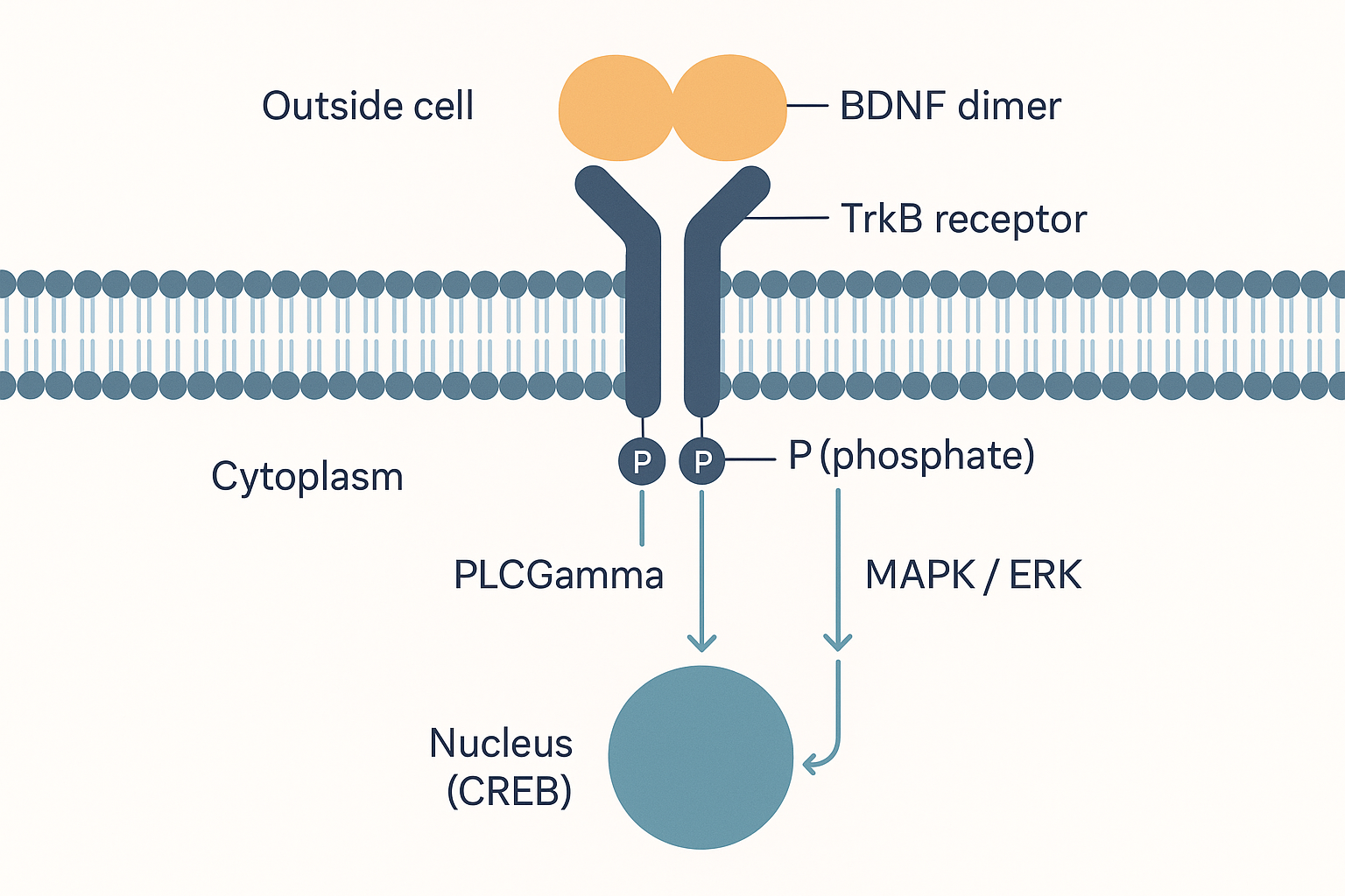 BDNF binds and pairs two TrkB receptors at the membrane, switching on their kinase tails. From there three cascades fan out — PLCγ to drive LTP now, PI3K/Akt to keep the cell alive, and MAPK/ERK to reach the nucleus and rewrite gene expression. This one docking event is what the entire class of "BDNF nootropics" is reaching for.
Activity-dependent release, exercise, and the Val66Met gene
Two facts make BDNF practically, not just theoretically, central.
First, BDNF release is activity-dependent. Neurons store BDNF in vesicles and release it when they fire — and the calcium that LTP lets in (via the NMDA receptor) also drives CREB to transcribe more BDNF. So activity makes BDNF, and BDNF enables the plasticity that activity is trying to produce: a virtuous loop. This is the molecular reason learning, novelty, and especially aerobic exercise raise BDNF — exercising muscle releases the protein irisin (FNDC5), which crosses into the hippocampus and induces BDNF transcription there, one of the cleanest mechanistic links between physical exercise and cognition. Conversely chronic stress, poor sleep, ageing, and a sedentary lifestyle lower BDNF, and low hippocampal BDNF is a core node of the neurotrophic hypothesis of depression (and part of why exercise and antidepressants, which both raise BDNF, help).
Second, there is a common genetic variant that throttles exactly this step: the BDNF Val66Met polymorphism. At position 66 of the pro-region, some people carry a methionine (Met) instead of a valine (Val). The Met version is poorly trafficked to the points of activity-dependent release — Met-BDNF "fails to localise to secretory granules and synapses" — so Met carriers secrete less BDNF in response to activity (their constitutive, baseline secretion is unchanged; it is the activity-triggered burst that suffers). The behavioural correlate, robust across studies, is poorer episodic memory and altered hippocampal function in Met carriers. The Met allele is common but very unevenly distributed geographically (rare in some African populations, up to ~40–50% allele frequency in parts of East Asia). It is a concrete, individual reason the same plasticity intervention lands differently in different people — some are starting from a lower activity-dependent BDNF ceiling.
Beyond the family: GDNF, CNTF, and IGF-1
Three further growth factors matter for the brain and are routinely (loosely) lumped under "neurotrophic", though strictly only the first four above are neurotrophins. The distinction is real: these three use completely different receptors, which is why they are not interchangeable.
GDNF (glial-cell-line-derived neurotrophic factor) is the most important of them and deserves its own paragraph. Despite the similar name it is not a neurotrophin and does not use a Trk receptor. It signals through a two-part receptor: a binding co-receptor, GFRα1, paired with the signalling kinase RET (a receptor tyrosine kinase). Its standout role is as the survival and trophic factor for midbrain dopaminergic neurons — the very neurons of the substantia nigra that die in Parkinson's disease (recall the nigrostriatal pathway from the cognition page). This made GDNF one of the most hoped-for drugs in neurology: directly infused into the putamen of Parkinson's patients it showed promise in early trials, but larger controlled trials were disappointing — defeated, like every trophic protein, by the delivery problem (a large protein that does not cross the blood-brain barrier and is hard to distribute evenly in tissue). GDNF also supports motor neurons and, outside the brain, is essential for kidney and enteric-nervous-system development.
CNTF (ciliary neurotrophic factor) signals through yet another system — a tripartite receptor sharing the gp130 subunit with inflammatory cytokines — and supports motor and other neurons; it also has notable effects on appetite and on hippocampal neurogenesis. Its practical relevance here is that P21 is a CNTF-derived peptide engineered to be small, stable, brain-penetrant and orally active — a worked example of the "shrink the trophic protein to a usable fragment" strategy.
IGF-1 (insulin-like growth factor 1) is a systemic growth hormone-driven factor (made largely in the liver) that also acts as a potent neurotrophic and neuroprotective signal in the brain via its own tyrosine-kinase receptor, overlapping heavily with the PI3K/Akt survival arm above. It is part of why metabolic health, exercise, and insulin signalling tie into brain plasticity — and why intranasal insulin appears in plasticity stacks.
Adult neurogenesis: the honest version
The flashiest plasticity claim is neurogenesis — that the adult brain grows brand-new neurons. It is real in some animals and some niches; in adult humans it is genuinely contested, and this database's job is to calibrate rather than cheerlead.
Where it happens (when it happens). Adult neurogenesis is not brain-wide. In adult mammals it is confined to (at most) two niches:
- The subgranular zone (SGZ) of the dentate gyrus, a sub-region of the hippocampus (the brain's memory-forming structure). Here resident neural stem cells divide, producing neuroblasts (immature migrating neurons) that mature into new granule neurons and wire into the existing hippocampal circuit. New, hyper-excitable young neurons are thought to aid pattern separation — telling similar memories apart — and this niche is the focus of nearly all the interest.
- The subventricular zone (SVZ) lining the ventricles, which in rodents feeds new neurons to the olfactory bulb. In adult humans this route appears largely redirected or absent after infancy.
What drives it (in the animal models where it is robust). The pro-neurogenic levers are strikingly the same healthy-brain levers as the rest of this page: aerobic exercise (the strongest), environmental enrichment and learning, good sleep, and BDNF. The suppressors are chronic stress and cortisol, inflammation (see the gut-brain axis — neuroinflammation suppresses both neurogenesis and BDNF), ageing, and poor metabolic health. This convergence is why "boost neurogenesis" and "boost BDNF / do cardio / sleep / lower inflammation" are, in practice, almost the same prescription.
The human controversy — calibrated. Whether meaningful neurogenesis continues in the adult human hippocampus is one of neuroscience's live arguments, and two landmark 2018 papers landed on opposite sides in the same months:
- Sorrells et al. (2018, Nature) found new-neuron markers dropping sharply in childhood and undetectable after ~13 years of age — concluding adult human hippocampal neurogenesis is essentially nil.
- Boldrini et al. (2018, Cell Stem Cell) examined hippocampi from people aged 14–79 and found thousands of immature neurons persisting into old age, with little decline.
- Moreno-Jiménez et al. (2019, Nature Medicine) found abundant immature neurons in healthy adults that declined sharply and early in Alzheimer's disease — and argued the disagreement is largely methodological (how the tissue is fixed and how long it sits before fixation dramatically affects whether the fragile markers survive).
The earlier, much-cited estimate that humans add roughly 700 new hippocampal neurons per day (Spalding et al., 2013, via carbon-14 birth-dating of cells) sits in the middle of this and is itself debated. The honest current reading: adult human hippocampal neurogenesis probably exists but at a low and uncertain rate, is highly sensitive to how it is measured, and almost certainly declines with age. It is a real phenomenon to support with the levers above — but any compound sold on a promise to flood your brain with new neurons is far ahead of the evidence. Most of what you can actually influence is the synaptic and structural plasticity of the neurons you already have.
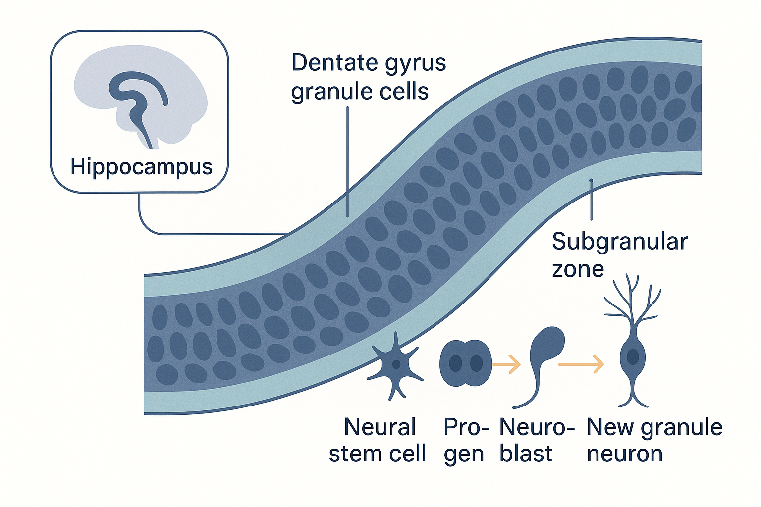 The one well-studied neurogenic niche: the subgranular zone of the hippocampal dentate gyrus. Resident neural stem cells give rise to neuroblasts that mature into new granule neurons and wire into the memory circuit — robust in rodents, real but low and contested in adult humans.
How the compounds engage these pathways
Now the payoff for the biohacker. Almost every "plasticity", "neuro-regeneration", or "smart-peptide" compound is reaching for one specific point on the map we have just built. Pinning each one to its level and molecule is what separates a mechanism from a marketing slogan — and several of these collapse to the same target dressed differently.
- Raising BDNF transcription / TrkB activation is by far the most crowded lever. Semax rapidly upregulates BDNF and TrkB phosphorylation in the hippocampus (a single intranasal dose raises BDNF mRNA ~3× in rodents). Noopept raises BDNF and NGF. Cerebrolysin and P21 are explicit attempts to mimic trophic-factor signalling with small BBB-crossing fragments (P21 is CNTF-derived and upregulates BDNF while inhibiting GSK3β). All of these are trying to switch on the BDNF → TrkB → CREB cascade described above — the difference is upstream (more BDNF released) versus the next two.
- Directly agonising the TrkB receptor (skipping BDNF entirely) is the cleaner version of the same idea. Eutropoflavin (4′-DMA-7,8-DHF) is a small-molecule TrkB agonist — a flavonoid that binds TrkB and triggers the same dimerisation-and-autophosphorylation cascade BDNF would, with the advantage of being a stable, BBB-penetrant molecule. NSI-189 is pitched at the neurogenesis/BDNF end of the same pathway.
- Synaptogenesis / structural plasticity is dihexa's headline claim — promoting new synapse formation (via an HGF/c-Met mechanism, not the neurotrophins) acting at the structural level (new spines/connections) rather than the synaptic-strength level. Read its deep dive for an unusually hard evidence-calibration: the famous "ten-million-times BDNF" potency claim rests partly on a retracted paper, and c-Met is an oncogene — a sharp reminder that pushing growth signalling is not free.
- GDNF / the dopaminergic arm. Bromantane is claimed in community use to raise GDNF and to upregulate dopamine synthesis (a different lever from the reuptake-blocking stimulants on the cognition page). Calibrate honestly: bromantane's well-supported action is on dopamine-synthesis enzymes; the GDNF link is weaker and the dopaminergic effects in the literature appear at doses well above typical use. It is better characterised as anxiolytic/anti-fatigue than as a proven neurotrophic agent.
- Tuning the trigger. Magnesium does not push growth at all — it tunes the NMDA magnesium block, keeping the coincidence detector's signal-to-noise crisp so that the right calcium signals get through. It acts at the very first step of the whole cascade.
The unifying caution, straight from the homeostatic-plasticity section: the brain defends a plasticity set-point, and crude, network-wide trophic pushes can degrade top-down control (several experienced users specifically flag rapid-plasticity agents like dihexa for disrupting prefrontal regulation). More BDNF everywhere is not obviously better; targeted plasticity is the goal, and the most reliable levers remain the unglamorous ones — exercise, sleep, learning, and low inflammation — that the molecules are all imitating.
Putting it all together
- Plasticity is a stack of four distinct levels: tuning a synapse's strength (synaptic), growing or pruning the hardware (structural), reassigning regional jobs (functional remapping), and birthing new cells (neurogenesis). Any "plasticity" claim is empty until pinned to a level.
- The glutamatergic synapse runs on two receptors. AMPA receptors carry the everyday sodium signal and their number is the synapse's strength; the NMDA receptor is a magnesium-plugged coincidence detector that opens its calcium gate only when glutamate and depolarisation occur together — the physical form of "fire together, wire together".
- Calcium is the instruction. A big fast NMDA calcium spike triggers LTP (CaMKII latches on → AMPA receptors phosphorylated and inserted → synapse louder → CREB → durable, structural); a small slow rise triggers LTD (phosphatases remove AMPA receptors → synapse weaker). Homeostatic scaling and metaplasticity defend a set-point so the system cannot run away.
- Structural plasticity writes the change into the dendritic spine: actin polymerises, a thin spine swells into a stable mushroom spine, and unused spines are pruned. Strength becomes shape.
- The neurotrophins gate all of it. Mature NGF/BDNF/NT-3/NT-4 → Trk receptors signal grow/survive/strengthen; pro-forms → p75 signal retract/prune/die. BDNF → TrkB fans into three cascades — PLCγ (LTP now), PI3K/Akt (survival), MAPK/ERK → CREB (durable, structural, and more BDNF). BDNF release is activity-dependent, is raised by exercise (via irisin) and lowered by stress/ageing, and is throttled in Val66Met Met carriers.
- GDNF (RET/GFRα, the dopaminergic-neuron trophic factor), CNTF, and IGF-1 sit alongside the family with different receptors; all trophic-protein drugs share one defeat — the blood-brain-barrier delivery problem.
- Adult human neurogenesis is confined to the hippocampal dentate gyrus, is driven by the same levers (exercise, sleep, BDNF, low inflammation), and is genuinely contested — probably real but low, age-declining, and measurement-sensitive. Be sceptical of "grow new neurons" promises; most of what you can move is the synaptic and structural plasticity of existing cells.
- Every plasticity compound targets one point on this map — and most cluster on BDNF/TrkB (Semax, Noopept, Cerebrolysin, P21, eutropoflavin), with dihexa at the structural/synaptogenic level, bromantane on the dopaminergic/GDNF arm, and magnesium tuning the NMDA trigger. The molecules are all imitating exercise, sleep, and learning — which remain the levers with the best evidence.
Related Compounds & Deep Dives
BDNF / TrkB pathway
- Semax & Selank — Semax rapidly upregulates hippocampal BDNF and TrkB phosphorylation; the canonical "BDNF peptide".
- Cerebrolysin — a standardised peptide hydrolysate engineered to mimic trophic-factor (BDNF/NGF/GDNF) signalling with BBB-crossing fragments.
- P21 (P021) — CNTF-derived, orally active peptide that upregulates BDNF and neurogenesis (inhibits GSK3β and LIF).
- Eutropoflavin (4′-DMA-7,8-DHF) — small-molecule TrkB agonist that mimics BDNF binding directly.
- Noopept — dipeptide nootropic that raises BDNF and NGF.
- NSI-189 — BDNF/neurogenesis-targeted antidepressant candidate.
Structural / synaptogenic plasticity
- Dihexa — claimed synaptogenic (new-synapse) agent via HGF/c-Met; read for the evidence-calibration on its hype (and a retracted key paper).
Dopaminergic / GDNF arm
- Bromantane — community-claimed GDNF and dopamine-synthesis upregulator; calibrated honestly (best-supported action is on dopamine synthesis, not proven neurotrophic effect).
Tuning the NMDA trigger
- Magnesium — keeps the NMDA-receptor magnesium block crisp as a signal-to-noise filter at the first step of the plasticity cascade.
Lifestyle lever
- Exercise — the strongest natural driver of BDNF (via muscle-derived irisin) and of hippocampal neurogenesis.
Related foundations
- Neuroscience of Cognition — the sibling page: dopamine synthesis and pathways, the synapse and reuptake, the prefrontal inverted-U.
- Cellular Energy — the glucose and ATP supply that powers the metabolically expensive work of plasticity.
- Gut-Brain Axis — how inflammation and LPS suppress BDNF, neurogenesis, and synaptic plasticity.