The Lymphatic System
The body's second circulation — the one with no heart. A one-way drainage network that mops up the few litres of fluid your blood vessels leak every day, doubles as the patrol-and-checkpoint system of your immune defence, ferries dietary fat into your bloodstream, and — discovered only in the last decade — even rinses metabolic waste out of your sleeping brain. Why movement, breathing, and sleep matter, and what the wellness world gets right and wrong about "lymphatic drainage".
The lymphatic system was glimpsed long before it was understood. In 1622 the Italian anatomist Gaspare Aselli, dissecting a well-fed dog, noticed milky-white threads running through the gut mesentery — he called them venae lactae, "milky veins". They were the lacteals, the lymphatic vessels that carry absorbed dietary fat, and they are the reason fresh chyle looks like milk (see Digestion). For two centuries the system stayed a curiosity. Then in 1896 the English physiologist Ernest Starling worked out the physics of why tissues need draining at all — the balance of pressures across capillary walls now called the Starling forces — and in doing so explained precisely what the lymphatics are for. The modern surprise came a century later still: in 2012 Maiken Nedergaard's group described the glymphatic system, a lymphatic-like waste-clearance route through brain tissue, and in 2015 Jonathan Kipnis and Antoine Louveau (with Aspelund and colleagues independently) rediscovered functional meningeal lymphatic vessels in the membranes around the brain — overturning the textbook dogma that the brain had no lymphatics at all. Current research is intense and still evolving: meningeal-lymphatic decline is now implicated in the build-up of amyloid-beta and tau in Alzheimer's disease, glymphatic clearance is found to run mostly during deep sleep, and in 2023 Nedergaard's group described a fourth meningeal membrane (the SLYM) — so treat the brain-clearance section as real and important but younger and less settled than the rest.
Why this page exists
The digestion page introduced "two highways out of the gut" — the portal vein and the lymphatic route — and promised the lymphatic side would get its own treatment. This is it. But fat transport, it turns out, is only the third most important thing the lymphatic system does.
The lymphatic system is the body's least-appreciated organ network, partly because it is almost invisible — its vessels are thin, clear, and easy to miss on dissection, and it has no thumping pump to draw attention to itself the way the heart does. Yet it solves a problem your cardiovascular system cannot solve on its own, it is the physical stage on which your immune system meets its enemies, and it has lately become one of the hottest topics in neuroscience. It also sits underneath a great deal of wellness marketing — "lymphatic drainage", "detox", dry-brushing, rebounding — some of which is physiologically sensible and some of which is overreach. This page builds the real system from the ground up, so that by the end you can tell which is which.
We start, as Starling did, with a leak.
The problem it solves: your blood vessels leak
Your smallest blood vessels, the capillaries (microscopic, thin-walled tubes where blood actually exchanges materials with tissue), are deliberately leaky. They have to be: oxygen, glucose, salts, and water must be able to pass out of the blood and into the tissue to feed the cells. The driving force is blood pressure — specifically the hydrostatic pressure (the physical push of fluid against the vessel wall) at the arterial end of the capillary, which squeezes plasma (the watery, protein-containing liquid part of blood, minus the cells) out through the wall into the spaces between cells.
That fluid, once it has left the blood and is bathing the tissue cells, has a new name: interstitial fluid (interstitial = "in the gaps", from the Latin for the spaces between things). It is essentially plasma minus most of the large proteins.
Now, why doesn't the tissue simply swell up forever? Because there is an opposing force pulling fluid back in. The blood retains large proteins — chiefly albumin — that are too big to leak out easily. Dissolved proteins draw water toward themselves by osmosis (water moving across a barrier toward the side with more dissolved particles); the resulting inward pull is called oncotic (or colloid osmotic) pressure. At the venous end of the capillary, where blood pressure has fallen, this inward oncotic pull wins, and most of the leaked fluid is sucked back into the blood. This tug-of-war between the outward push of hydrostatic pressure and the inward pull of oncotic pressure is exactly what Starling formalised — the Starling forces.
Here is the crucial arithmetic. The filtration out and the reabsorption back do not balance. Across the whole body, capillaries push out roughly 20 litres of fluid per day, and reabsorb only about 17 litres. That leaves around 2–3 litres of interstitial fluid stranded in the tissues every single day — plus the small amount of protein that inevitably escapes the blood and, being large, cannot be reabsorbed by osmosis at all.
If that 2–3 litres simply accumulated, you would swell visibly within hours and be dead within days. It must be collected and returned to the bloodstream. That return service is the lymphatic system. It is a one-way drainage network that picks up the leftover interstitial fluid (and the stranded protein), and pipes it back to the great veins near the heart. Once interstitial fluid enters a lymphatic vessel, we give it yet another name: lymph. Lymph is nothing exotic — it is simply the body's leaked fluid, on its way home.
flowchart LR
ART["Arterial end of capillary<br/>high blood pressure"] -->|"pushes ~20 L/day OUT<br/>(hydrostatic)"| TISS["Interstitial fluid<br/>(bathes the cells)"]
TISS -->|"oncotic pull sucks<br/>~17 L/day back IN"| VEN["Venous end of capillary"]
TISS -->|"leftover ~2-3 L/day<br/>+ stranded protein"| LYMPH["Lymphatic capillary<br/>(fluid now = LYMPH)"]
LYMPH -->|"one-way return"| SUB["Subclavian vein<br/>back to bloodstream"]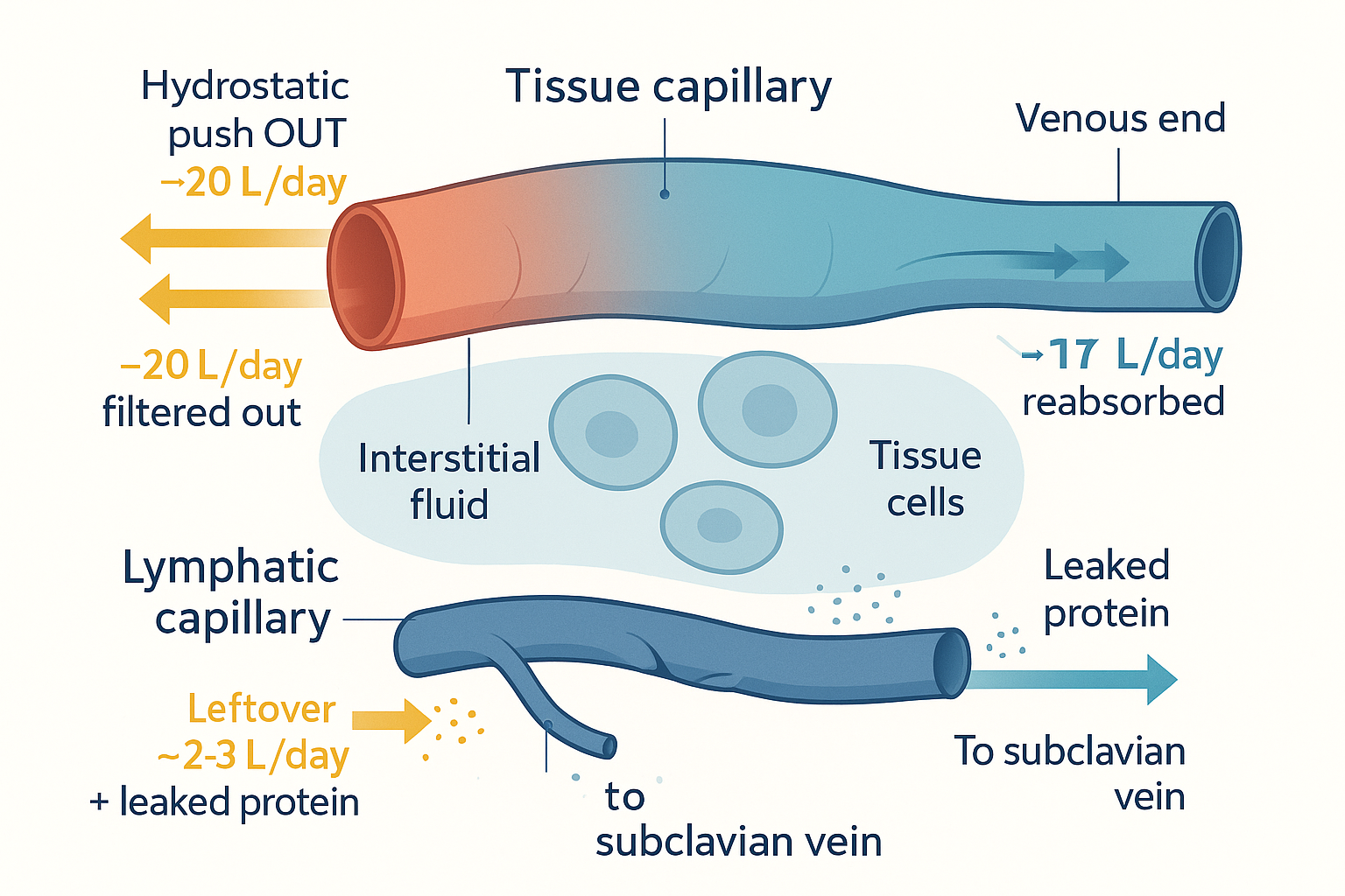 The leak and its remedy: capillaries filter ~20 L/day out and reabsorb ~17 L; the lymphatic capillary collects the leftover ~2–3 L (plus protein osmosis cannot reclaim) and returns it to the bloodstream.
This reframes the whole system. The lymphatic network is not a mysterious "detox" organ — it is, first and foremost, the overflow drain that keeps your tissues from waterlogging and recovers the protein your blood would otherwise lose. Everything else it does is built on top of that drainage plumbing.
Architecture and flow: a network with no pump
Trace the journey of a litre of lymph from tissue to bloodstream and the architecture reveals itself as a series of progressively larger vessels, punctuated by filtration stations.
1. Lymphatic capillaries — the inlets. These are the smallest, most peripheral lymphatic vessels, and they are blind-ended — they begin as closed tubes out in the tissue, like the dead-end fingers of a glove, with no upstream connection. Their walls are a single layer of endothelial cells (the flat cells that line all vessels) arranged in an ingenious way: the cells overlap like roof shingles or loose flaps, anchored to the surrounding tissue by tiny filaments. When interstitial fluid accumulates and tissue pressure rises, it pushes these flaps inward — they swing open like little one-way doors, letting fluid (and particles far too large for a blood capillary, including whole proteins, bacteria, and immune cells) flood in. Once fluid is inside and pressure equalises, the flaps fall shut, so fluid cannot leak back out. These overlapping flaps are the system's primary valves, and they are the reason the lymphatic capillary is such an effective one-way inlet: it can swallow large debris that the blood capillaries refuse, and it never gives it back.
2. Collecting lymphatic vessels — the pumps. Lymphatic capillaries merge into larger collecting vessels, and here the design changes in two important ways. First, these vessels are studded with secondary valves — proper one-way valves, like the bicuspid valves in your veins, spaced along the vessel so lymph can only travel forward. Second, the vessel wall now contains a layer of smooth muscle (involuntary muscle, the kind that lines your gut and arteries). The segment of vessel between two consecutive valves — a muscular chamber bracketed by one-way doors — is called a lymphangion, and it is the closest thing the lymphatic system has to a heart. Each lymphangion contracts rhythmically, squeezing its lymph through the downstream valve into the next lymphangion, which then contracts in turn — a travelling wave of squeezes marching lymph forward. A chain of lymphangions is, in effect, a string of tiny serial hearts.
3. Lymph nodes — the checkpoints. Before lymph completes its journey it is forced through a series of lymph nodes — small, bean-shaped filtration-and-immune stations (covered in detail below). This is where the drainage system doubles as an immune-surveillance system.
4. Trunks and ducts — the outlets. Collecting vessels merge into larger lymphatic trunks, which in turn drain into one of two great ducts. The thoracic duct is the main one — a vessel running up the back of the chest that drains the entire lower body, the abdomen, the left arm, and the left side of the head and chest: roughly three-quarters of the body. The smaller right lymphatic duct drains the right arm and the right side of the head and chest. Both empty into the subclavian veins (the large veins beneath the collarbones, sub-clavian = "under the clavicle"), at the junction where they meet the internal jugular veins — returning the lymph to the bloodstream and closing the loop that began with the capillary leak.
flowchart LR
CAP["Interstitial fluid"] -->|"flaps swing in<br/>(primary valves)"| LC["Lymphatic capillary<br/>blind-ended, overlapping flaps"]
LC --> COLL["Collecting vessel<br/>lymphangions: smooth muscle<br/>+ one-way (secondary) valves"]
COLL --> NODE["Lymph node<br/>(filter + immune checkpoint)"]
NODE --> TRUNK["Lymphatic trunks"]
TRUNK --> TD["Thoracic duct (≈3/4 of body)<br/>+ right lymphatic duct"]
TD --> SCV["Subclavian veins<br/>→ bloodstream"]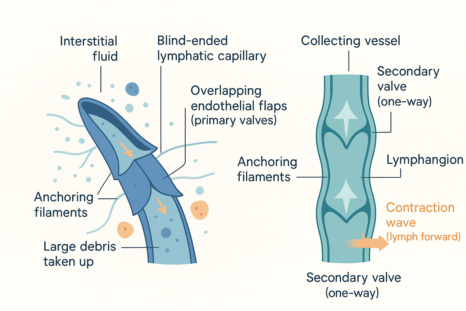 The one-way machine: overlapping flaps (primary valves) let fluid and large debris into the blind-ended capillary; downstream, smooth-muscle lymphangions bracketed by secondary valves actively pump lymph forward.
Now the single most important fact about lymphatic flow, and the one that underlies almost every practical recommendation on this page: there is no central pump. The heart drives blood around a closed loop, but the lymphatic system is an open-ended, low-pressure network, and it has nothing equivalent to the heart. So what moves the lymph? Four forces, working together:
- Skeletal-muscle pumping. This is the big one. When the muscles around a lymphatic vessel contract — every time you walk, fidget, or flex — they squeeze the vessel, and because the secondary valves only allow forward flow, each squeeze ratchets lymph onward. Your calf muscles in particular act as a powerful "peripheral pump" for the lymph (and venous blood) of the legs. This is the mechanistic reason physical movement genuinely aids lymph flow, and why prolonged immobility — long flights, bed rest — causes the ankles to swell.
- The respiratory pump. Breathing changes the pressures in the chest and abdomen. Each inhalation drops the pressure in the thoracic cavity and raises it in the abdomen, creating a pressure gradient that effectively sucks lymph up the thoracic duct toward the chest. Deep, slow breathing measurably assists lymphatic return — the same diaphragmatic breathing that raises vagal tone on the autonomic nervous system page.
- Arterial pulsation. Lymphatic vessels often run alongside arteries, and the pulse of the artery with each heartbeat massages the adjacent lymphatic, helping to milk it forward.
- Intrinsic lymphangion contraction. As described above, the lymphangions contract on their own, autonomously, like little hearts — and they pump faster when stretched by a greater volume of lymph (a self-regulating response). This intrinsic pumping is the system's baseline, working even at rest, but it is greatly amplified by the external forces above.
The takeaway: because lymph depends on muscle contraction, breathing, and movement rather than a dedicated pump, a sedentary, shallow-breathing body genuinely drains its tissues more sluggishly — and conversely, movement and breathing are the legitimate, mechanistically sound core of "getting the lymph moving". Hold this; it is the honest kernel inside the wellness claims we will scrutinise later.
The three jobs
With the plumbing established, the lymphatic system's three functions fall out naturally. All three ride on the same drainage network.
flowchart TD
    LYMPH["LYMPHATIC SYSTEM<br/>(one drainage network)"]
    LYMPH --> J1["1. FLUID HOMEOSTASIS<br/>return leaked fluid + protein<br/>to the bloodstream"]
    LYMPH --> J2["2. IMMUNE SURVEILLANCE<br/>lymph nodes sample lymph;<br/>antigen meets lymphocyte"]
    LYMPH --> J3["3. DIETARY FAT TRANSPORT<br/>lacteals carry chylomicrons<br/>into blood (bypass liver)"]
Job 1: Fluid homeostasis (the drainage we have already met)
This is the foundational job, covered above: collect the ~2–3 L/day of leaked interstitial fluid and the escaped protein, and return them to the blood. When this fails, fluid pools in the tissues — oedema (swelling). Keep this in mind; it is the failure mode we return to at the end.
Job 2: Immune surveillance — the lymph node as a meeting hall
The lymphatic system does not merely transport fluid; it transports a sample of everything happening in your tissues — and it routes that sample through inspection stations. This makes it the backbone of the adaptive immune system (the part of immunity that learns to recognise specific threats; covered fully on the forthcoming Immune System foundations page).
Consider what the lymph carries. Because the lymphatic capillaries take up large particles that blood capillaries reject, lymph draining from an infected or injured tissue is laden with antigens (molecular fragments of pathogens or damaged cells — literally anything the immune system might need to recognise), often physically carried by dendritic cells (sentinel immune cells whose job is to grab antigen in the tissues and carry it to where it can be shown to the rest of the immune system). All of this flows toward the lymph nodes.
A lymph node is a small encapsulated organ, a few millimetres across, positioned along the lymphatic vessels — clustered especially in the neck, armpits, groin, and gut mesentery (the places where you "feel your glands swell" when ill; that swelling is a node working overtime). Lymph enters a node through several afferent vessels (afferent = "carrying toward"), percolates slowly through a maze of internal channels — being filtered the whole way — and exits, cleaned and inspected, through a single efferent vessel (efferent = "carrying away"). Inside, the node is rigorously organised so that the right immune cells meet the right cargo:
- The cortex (outer region) is packed with B-cell follicles — dense clusters of B lymphocytes (the immune cells that produce antibodies, the targeting proteins that lock onto specific antigens).
- The paracortex (the layer just beneath) is the T-cell zone, rich in T lymphocytes (the immune cells that coordinate the response and kill infected cells). This is where dendritic cells, having arrived from the tissue, present their captured antigen to T cells — the pivotal molecular handshake that activates the adaptive response.
- The medulla (inner region) contains the cells that secrete finished antibody into the outgoing lymph.
So the lymph node is a meeting hall: it concentrates incoming antigen in one place and parades it past a dense, waiting population of lymphocytes, so that the rare lymphocyte capable of recognising that specific threat is far more likely to encounter it. This is why nodes swell during infection (lymphocytes proliferating en masse), and — soberingly — why cancers metastasise via the lymphatics: the same network that samples your tissues also offers tumour cells a highway to spread, which is why surgeons examine and sometimes remove the "sentinel" lymph node draining a tumour.
flowchart LR
TISSUE["Infected tissue<br/>antigen + dendritic cells"] -->|afferent vessels| NODE
subgraph NODE["LYMPH NODE"]
CORTEX["Cortex<br/>B-cell follicles<br/>(antibody producers)"]
PARA["Paracortex<br/>T-cell zone<br/>(dendritic cell presents antigen)"]
MED["Medulla<br/>(antibody secretion)"]
end
NODE -->|efferent vessel| ONWARD["Cleaned lymph<br/>+ activated lymphocytes,<br/>antibody → bloodstream"]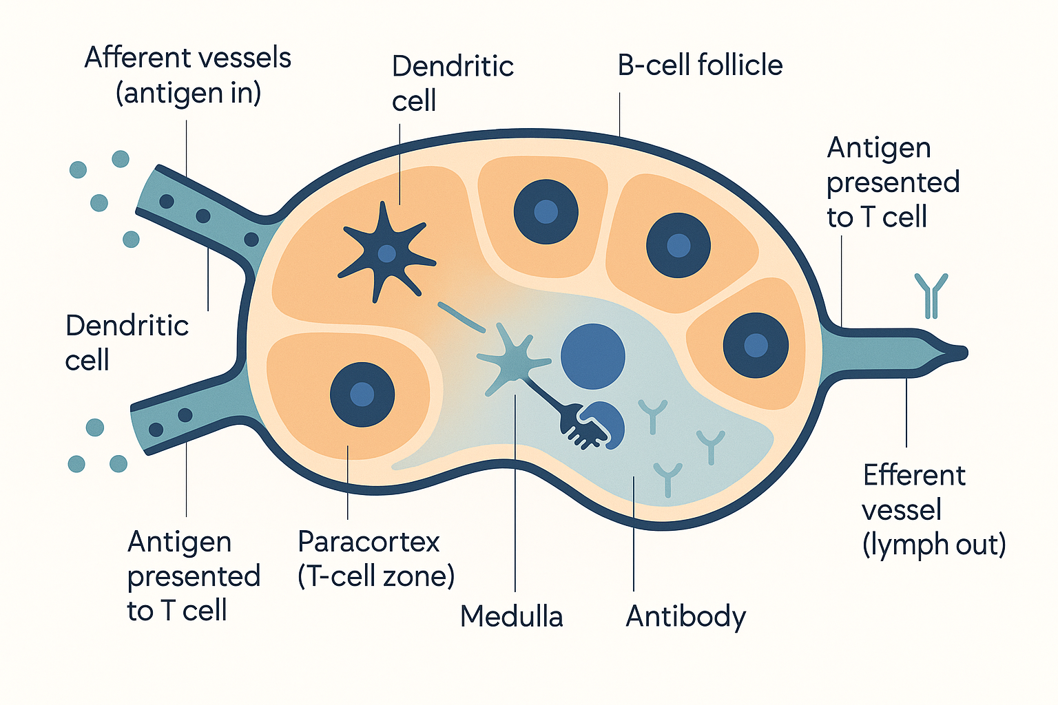 The immune checkpoint: afferent vessels deliver antigen-laden lymph into the node, where dendritic cells present antigen to T cells in the paracortex and B-cell follicles in the cortex make antibody; filtered lymph and activated cells leave by the efferent vessel.
The lymph nodes are the most numerous immune organs, but they have relatives that work on the same principle of "concentrate the lymphocytes where the threats arrive":
- The spleen does for the blood what lymph nodes do for the lymph — it filters blood, traps blood-borne pathogens, and houses a large reserve of lymphocytes (it is also the hub of the vagal anti-inflammatory reflex from the autonomic nervous system page).
- The tonsils guard the entrance to the throat.
- MALT and GALT — mucosa-associated and gut-associated lymphoid tissue — are patches of immune tissue stationed directly under the linings of the gut and airways, where the outside world presses closest against the body. GALT alone holds a large fraction of all your immune cells, because the gut is the body's biggest interface with foreign material (a recurring theme on the gut microbiome page). The thymus, where T cells are trained, is the other primary lymphoid organ — it gets its own forthcoming Thymus page.
Job 3: Dietary fat transport — the lacteals
The third job is the one the digestion page already opened: the lymphatic system is the route by which dietary fat enters the bloodstream. We will not repeat that page's detail — only connect it to the architecture just described.
Recall that each finger-like villus lining the small intestine contains, at its core, a single blind-ended lymphatic capillary called a lacteal (Aselli's "milky vein"). When you digest fat, the intestinal cells reassemble it into large fat-and-protein particles called chylomicrons — too big to squeeze into a blood capillary. But they fit easily through the generous, flap-valved opening of a lacteal. So fat-laden chylomicrons enter the lymphatic system, turning the lymph draining the gut milky-white (this fluid is specifically called chyle). That chyle flows up through the lymphatic trunks, into the thoracic duct, and empties into the subclavian vein — depositing dietary fat directly into the bloodstream and thereby bypassing the liver's first-pass metabolism, exactly the "lymphatic highway" the digestion page contrasted with the portal vein.
Two practical consequences, both already flagged on the digestion page and worth restating in lymphatic terms:
- The fat-soluble vitamins A, D, E, and K ride this same lymphatic route (they dissolve into the chylomicrons), which is why they need dietary fat for absorption and have a slow, delayed onset — they take the long lymphatic detour.
- Medium-chain fats (MCTs / coconut oil) are the exception: small and water-soluble enough to skip the chylomicron-and-lacteal route entirely and take the fast portal vein to the liver — the reason MCTs are prized as a "fast" fat fuel.
This is also why a leak or blockage of the thoracic duct (from surgery or injury) can produce chylothorax — milky, fat-rich lymph leaking into the chest cavity — a vivid demonstration that your dinner's fat really does travel by this route.
The glymphatic system: the brain's lymphatic-like clearance
For over a century, anatomy textbooks stated flatly that the brain has no lymphatic vessels. This was a genuine puzzle, because the brain is the body's most metabolically active organ — it generates a great deal of waste — and every other tissue relies on lymphatics to clear leaked protein and debris. How could the one organ that most needs waste clearance have no drainage system? The answer, worked out only since 2012, is that the brain has its own clearance arrangement, lymphatic in spirit if not in exact anatomy. This is the glymphatic system — a portmanteau of "glial" and "lymphatic", because glial cells (the brain's non-neuronal support cells) do the job that lymphatic vessels do elsewhere.
Here is how it works. The brain floats in cerebrospinal fluid (CSF) — the clear fluid that cushions it. The glymphatic insight is that CSF does not just sit around the brain; it is actively driven through the brain tissue to flush it. The route is anatomically specific: CSF is pumped into the depths of the brain along the outsides of the arteries — the perivascular spaces (peri-vascular = "around the vessels"), narrow sleeves of fluid wrapping every penetrating blood vessel. To get from those sleeves into the brain tissue proper, the fluid passes through channels formed by astrocytes (a star-shaped type of glial cell) whose end-feet wrap the vessels. These astrocyte end-feet are studded with a water channel protein called aquaporin-4 (AQP4), which is the gateway that lets CSF flood into the tissue. The incoming CSF then sweeps through the brain's interstitial spaces, mixes with the interstitial fluid bathing the neurons, picks up dissolved metabolic waste, and is flushed out along the veins — carrying the waste away, ultimately toward the genuine lymphatic vessels in the meninges (described next) and on to the lymph nodes of the neck.
The waste this clears is not trivial. It includes amyloid-beta and tau — the two proteins that aggregate into the plaques and tangles of Alzheimer's disease. The glymphatic system is, in effect, the brain's nightly rinse cycle for exactly the proteins implicated in neurodegeneration.
And "nightly" is precise, because the headline finding is this: glymphatic clearance runs predominantly during sleep, especially deep (slow-wave) sleep. During deep sleep, the spaces between brain cells widen substantially, dramatically increasing the flow of CSF through the tissue and accelerating waste removal. The brain, in other words, cleans itself while you sleep — and this is one of the most compelling mechanistic reasons yet found for why sleep is non-negotiable, and why chronic sleep deprivation may, over years, allow neurotoxic waste to accumulate. It connects sleep, the circadian rhythm, and possibly melatonin directly to long-term brain health.
flowchart LR
CSF["Cerebrospinal fluid (CSF)"] -->|"along arteries<br/>(perivascular space)"| AQP["Astrocyte end-feet<br/>AQP4 water channels"]
AQP --> TISSUE["Brain tissue<br/>CSF mixes with interstitial fluid"]
TISSUE -->|"picks up amyloid-β, tau"| OUT["Outflow along veins"]
OUT --> MENING["Meningeal lymphatic vessels"]
MENING --> NODES["Deep cervical lymph nodes (neck)"]
SLEEP["DEEP SLEEP<br/>cell spaces widen<br/>→ flow surges"] -.->|amplifies whole loop| TISSUE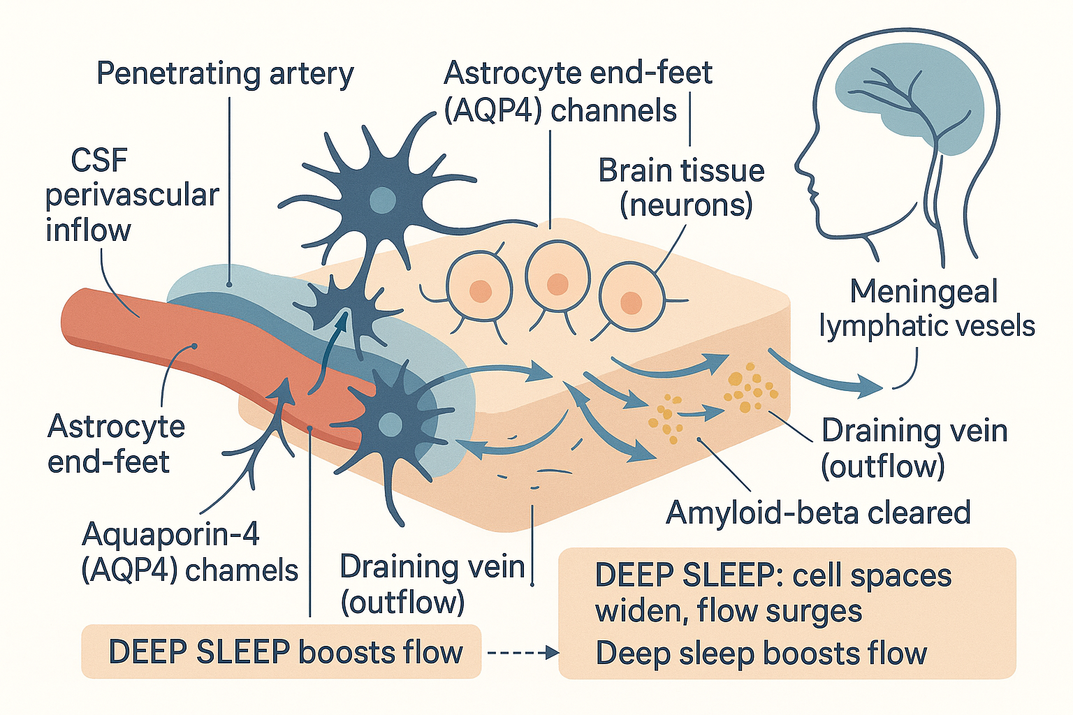 The brain's rinse cycle: CSF enters along arteries through AQP4 astrocyte channels, flushes interstitial waste (amyloid-beta, tau) out along veins to meningeal lymphatics and neck nodes — and the flow surges during deep sleep as the spaces between cells widen.
The second piece of the story is the meningeal lymphatic vessels. The meninges are the three protective membranes wrapping the brain and spinal cord. In 2015 two groups independently showed that these membranes do contain genuine lymphatic vessels — real ones, lined with the same markers as lymphatics elsewhere — running alongside the great venous sinuses and draining to the deep cervical lymph nodes in the neck. This was the missing downstream plumbing: the glymphatic system flushes waste out of the brain tissue, and the meningeal lymphatics carry it the rest of the way to the conventional lymphatic system. The two together form a complete brain-clearance pathway. Crucially, this also means the brain is not the "immune-privileged", immunologically sealed box it was long thought to be — it is in direct lymphatic communication with the immune system, with large implications for how we understand neuroinflammation, multiple sclerosis, and brain infection.
A calibration, as promised in the historical note: this is real, replicated, and important science, but it is young. The exact anatomy is still being refined (the 2023 description of the SLYM, a previously unrecognised fourth meningeal membrane, shows how fast the picture is still changing), the precise quantitative contribution of glymphatic flow in the human brain is debated, and the causal links to Alzheimer's disease — while strongly suggestive in animal models, where destroying meningeal lymphatics accelerates amyloid build-up — are not yet settled in humans. Treat it as a frontier worth knowing, not a closed textbook chapter. What is robust enough to act on today is the simplest implication: deep sleep drives brain waste clearance, so protecting sleep is protecting the rinse cycle.
Failure modes and "lymphatic drainage": the honest version
When the drainage job (Job 1) fails, the result is lymphoedema — chronic, often disfiguring swelling of a body part as protein-rich interstitial fluid accumulates because the lymph cannot carry it away. It comes in two forms:
- Primary lymphoedema is congenital — the person is born with malformed or insufficient lymphatic vessels (sometimes from identifiable gene mutations affecting lymphatic development).
- Secondary lymphoedema is acquired — the lymphatic system was working but got damaged or blocked. The two leading causes worldwide are illuminating. In high-income countries, the commonest cause is cancer treatment: removing or irradiating lymph nodes (e.g. the axillary nodes in breast-cancer surgery) severs the drainage route, and the arm or leg downstream swells — sometimes years later. Globally, the commonest cause is filariasis, infection by parasitic worms (Wuchereria bancrofti) that physically lodge in and block the lymphatic vessels, producing the gross swelling historically called elephantiasis. Both illustrate the same principle: break the one-way drain, and the tissue it served waterlogs with protein-rich fluid.
How is lymphoedema actually managed? The evidence-based mainstays are mechanical, and they map exactly onto the flow physics established earlier — there is no pump, so you assist flow externally:
- Manual lymphatic drainage (MLD) — a specific, gentle, light-pressure massage technique that physically milks lymph along its vessels toward functioning nodes. Note: this is gentle and directional, working with the low-pressure, valved architecture — not deep tissue kneading.
- Compression — bandaging or compression garments that raise tissue pressure and support the skeletal-muscle pump, the single most effective long-term measure.
- Exercise and movement — engaging the skeletal-muscle pump to drive flow (exercise, walking/running).
- Meticulous skin care — because stagnant, protein-rich lymph is a breeding ground for infection.
Notice that these are real medical interventions for a real failure of drainage in people whose lymphatics are damaged. This is where we must carefully separate the medicine from the marketing, because the wellness world borrows the term "lymphatic drainage" and applies it to healthy people with claims that range from physiologically sound to outright overreach.
What is genuinely, mechanistically plausible (and supported by the flow physics on this page):
- Movement and exercise aid lymph flow. Unambiguously true — the skeletal-muscle pump is a primary driver of lymph, and it is why immobility causes swelling and activity relieves it.
- Deep, slow breathing assists lymphatic return. True — the respiratory pump genuinely helps draw lymph up the thoracic duct.
- The calf-muscle pump matters, and elevating the legs or moving them after prolonged sitting genuinely reduces fluid pooling. True.
- Adequate hydration keeps lymph (which is mostly water) appropriately fluid; severe dehydration does not help drainage. Plausible and harmless.
- Manual lymphatic drainage helps people with actual lymphoedema. True, and evidence-based — in that population.
What is overreach or unproven when sold to healthy people:
- "Detox" framing. The lymphatic system returns leaked fluid and surveils for pathogens; it is not a toxin-elimination organ. The organs that actually clear metabolic and chemical waste are the liver (Phase I/II metabolism) and the kidneys. A "lymphatic detox" does not exist in the physiological sense the marketing implies — there is no reservoir of "toxins" stored in the lymph waiting to be flushed.
- Dry brushing and lymphatic-drainage massage "boosting detox" in healthy people. Gentle massage can transiently nudge lymph along, and may feel pleasant, but there is no good evidence it removes "toxins" or produces lasting health benefits in people with normal lymphatic function. The claimed mechanism is borrowed from MLD, which was designed for damaged lymphatic systems, not healthy ones.
- Rebounding (mini-trampoline) as a uniquely powerful "lymph flush". The kernel of truth is real — bouncing is movement, and movement drives lymph via the muscle pump — but there is nothing special about a trampoline versus walking, and claims of dramatic, quantified "lymph flushing" are unsupported. It is exercise, marketed as magic.
The fair summary: the lymphatic system genuinely responds to movement, breathing, posture, and (in the damaged case) skilled massage and compression — that part is real physiology. What is not real is the idea of the lymph as a "toxin sink" that special techniques uniquely "drain". The honest, useful version of "lymphatic health" is unglamorous and free: move your body, breathe deeply, sleep well, and don't sit immobile for hours. Those work because of exactly the no-pump architecture this page laid out.
Putting it all together
- The lymphatic system exists because blood capillaries leak. Starling forces push ~20 L/day of plasma into the tissues and reabsorb only ~17 L, stranding 2–3 L of interstitial fluid plus escaped protein that must be returned to the blood. The lymphatic system is that one-way return drain; once interstitial fluid enters it, the fluid is called lymph.
- Its architecture is a series of widening, valved vessels: blind-ended lymphatic capillaries (overlapping endothelial flaps as primary valves let fluid and large debris in) → collecting vessels with lymphangions (smooth-muscle chambers between secondary valves) → lymph nodes → trunks → the thoracic duct (≈¾ of the body) and right lymphatic duct → the subclavian veins.
- There is no central pump. Lymph moves by the skeletal-muscle pump, the respiratory pump, arterial pulsation, and intrinsic lymphangion contraction — which is exactly why movement, breathing, and not sitting immobile genuinely matter for lymph flow.
- It does three jobs on one network: (1) fluid homeostasis — return leaked fluid and protein; (2) immune surveillance — lymph nodes filter lymph and stage the antigen-meets-lymphocyte handshake (cortex/B-cells, paracortex/T-cells, medulla), alongside the spleen, tonsils, and gut-associated lymphoid tissue; (3) dietary fat transport — intestinal lacteals carry chylomicrons into the blood, bypassing the liver.
- The brain has its own version: the glymphatic system flushes CSF through brain tissue along perivascular AQP4 astrocyte routes, clearing amyloid-beta and tau, and it runs predominantly during deep sleep — draining via the rediscovered meningeal lymphatics to the neck nodes. Real and important, but younger and less settled than the rest.
- When drainage fails — lymphoedema (congenital, post-node-removal, or filariasis) — it is managed mechanically (manual lymphatic drainage, compression, exercise), consistent with the no-pump physics. The wellness "lymphatic detox" framing is largely overreach: movement, breathing, sleep, and hydration genuinely aid lymph; "flushing toxins" is marketing — the liver and kidneys, not the lymph, clear waste.
The unifying idea: the lymphatic system is the body's second circulation — a heartless, one-way drain that turns out to also be the stage for immune defence, the conduit for dietary fat, and (in the brain) the night-shift cleaner. Almost every legitimate way to "support" it comes down to supplying the pumping it lacks — through muscle, breath, and sleep.
Related Compounds & Deep Dives
Movement & the muscle pump (drive lymph flow)
- Exercise and running — the skeletal-muscle pump is a primary driver of lymph; movement is the most legitimate "lymphatic support" there is.
Sleep & the glymphatic rinse cycle
- Sleep — deep slow-wave sleep is when glymphatic brain-waste clearance surges; the strongest practical lever on the system.
- Melatonin and circadian rhythm — set the sleep timing that gates glymphatic clearance.
- Meditation — slow breathing assists the respiratory pump and lymphatic return, and supports sleep quality.
Dietary fat transport (the lacteal/chylomicron route)
- Coconut oil / MCTs — the exception that skips the lacteal/lymphatic route and takes the fast portal vein, illuminating the rule.
- ADEK fat-soluble vitamins, vitamin D, vitamin E, vitamin K — absorbed into chylomicrons and carried by the lymphatic highway, hence the need for dietary fat and the slow onset.
Immune / lymphoid tissue support
- Lactoferrin and colostrum — support mucosal (gut-associated) immune tissue; see the Lactoferrin & Colostrum deep dive.
- Vitamin D — broadly immunomodulatory across the lymphoid system.
Related foundations
- Digestion — the "two highways" (portal vein vs lymphatic) and the full chylomicron/lacteal fat-absorption story this page builds on.
- The Liver — the first-pass checkpoint the lymphatic fat route bypasses, and the organ that actually clears toxins (not the lymph).
- Inflammation — the immune response the lymph nodes stage, and the neuroinflammation the meningeal lymphatics now connect to the brain.
- Autonomic Nervous System — the respiratory/diaphragmatic pump and the spleen's vagal anti-inflammatory reflex.
- Gut Microbiome — the gut-associated lymphoid tissue (GALT) that holds much of the body's immune cell population.
- Immune System (forthcoming) — the adaptive immunity for which lymph nodes are the staging ground.
- Thymus (forthcoming) — the primary lymphoid organ where T cells are trained before patrolling the lymphatics.