The Pineal Gland
The tiny midline gland that turns darkness into a hormone — how the eye, the brain's master clock, and a single sympathetic nerve conspire to make melatonin at night, why that signal is also a potent antioxidant, and why the gland fills with "brain sand" as you age.
A short history first, because the pineal has the strangest reputation of any organ. In the 1640s the philosopher René Descartes singled it out as the "seat of the soul" — the unique unpaired point where, he reasoned, the immaterial mind met the physical body. He was wrong about the soul but right that the gland is peculiar: it is one of the few midline, unpaired structures in a brain otherwise built in mirror-image pairs. For three centuries it remained a mystery with no known function. Then in 1958, the dermatologist Aaron Lerner, hunting for a factor that lightened frog skin, isolated a pineal compound and named it melatonin (because it aggregated melanocyte pigment). In the 1960s, Julius Axelrod (later a Nobel laureate) worked out that the pineal makes melatonin on a daily rhythm controlled by light and the sympathetic nervous system — turning the "seat of the soul" into the body's measurable clock hormone. The final pieces came around 2000–2002, when researchers identified the suprachiasmatic nucleus as the brain's master clock and discovered a previously unknown class of light-sensing cells in the retina (the melanopsin-containing ganglion cells) that tell that clock whether it is day or night. Current research now studies melatonin's role in mitochondrial protection, the pineal's calcification and decline in ageing, and disturbed melatonin signalling in neurodegeneration — the threads this page pulls together.
Why this page exists
Most people meet the pineal gland only through a bottle of melatonin, and assume melatonin is simply "a sleep supplement." Both ideas badly undersell what is actually happening. The pineal is the hardware that converts a light signal into a chemical one — it is where the abstract fact "it is now night" becomes a molecule released into your blood, read by every organ. To understand it you have to follow a remarkable chain that begins in the eye, passes through the brain's master clock, descends into the spinal cord, loops back up through a nerve in your neck, and finally lands on the gland as a squirt of noradrenaline. That chain is one of the most elegant pieces of wiring in the body, and it explains everything downstream: why melatonin is made only in darkness, why blue light at night wrecks sleep, why jet lag happens, and why the hormone is also, almost as a side job, one of the most effective antioxidants your cells possess.
This page builds that system from the gland outward. It is the endocrine counterpart to the timing problem: where the hormones page covered what a hormone is and listed the pineal as one of the endocrine glands, and the autonomic nervous system page covered the sympathetic wiring that turns out to drive this gland, this page shows how those two systems meet in one organ to keep your biology on a 24-hour schedule. We finish on the two questions people actually ask about the pineal: why it calcifies with age (the "brain sand," and the fluoride argument — calibrated honestly), and whether the pineal peptides (Epitalon, Pinealon) can restore a fading gland.
Anatomy: a small gland in a strategic place
The pineal gland (also called the epiphysis cerebri) is tiny — roughly 5 to 8 mm long and weighing only about 100 to 150 mg, the size of a grain of rice. Despite that, it sits in one of the most central locations in the brain: on the midline, at the back of the third ventricle (one of the brain's fluid-filled chambers), in a region called the epithalamus (the "upper thalamus," just above and behind the thalamus, the brain's great sensory relay station). It is attached by a short stalk and tucked beneath the back end of the corpus callosum. Because the brain is otherwise built as two mirror-image halves, the pineal's position as a single, unpaired, midline organ is genuinely unusual — and is part of what gave it its mystical reputation.
Three anatomical facts about it matter mechanistically:
- Its cells. The pineal is built mostly (over 95%) of pinealocytes — specialised secretory cells, evolutionarily related to the light-sensing photoreceptors of the retina, that are the factories for melatonin. (In some non-mammalian vertebrates the pineal is directly light-sensitive — a literal "third eye." In mammals it lost direct photosensitivity and instead receives light information indirectly, through the nervous wiring described below.) The rest is supporting glial cells.
- It lies outside the blood-brain barrier. The blood-brain barrier (BBB) is the tight seal that normally walls the brain off from the general circulation, letting only selected molecules through. The pineal is one of a small number of brain structures that sit outside this barrier (a circumventricular organ). This has two consequences: the gland can dump melatonin straight into the bloodstream (and the cerebrospinal fluid) without having to cross the barrier, so its hormone reaches the whole body quickly; and, less happily, the gland is exposed to whatever circulates in the blood — including minerals, calcium, and fluoride, which matters for the calcification story later.
- Its blood supply is enormous for its size. Per gram of tissue, the pineal is one of the most richly perfused organs in the body, second only to the kidney. A gland whose entire job is to broadcast a hormone needs the plumbing to release it fast and widely — and it has it.
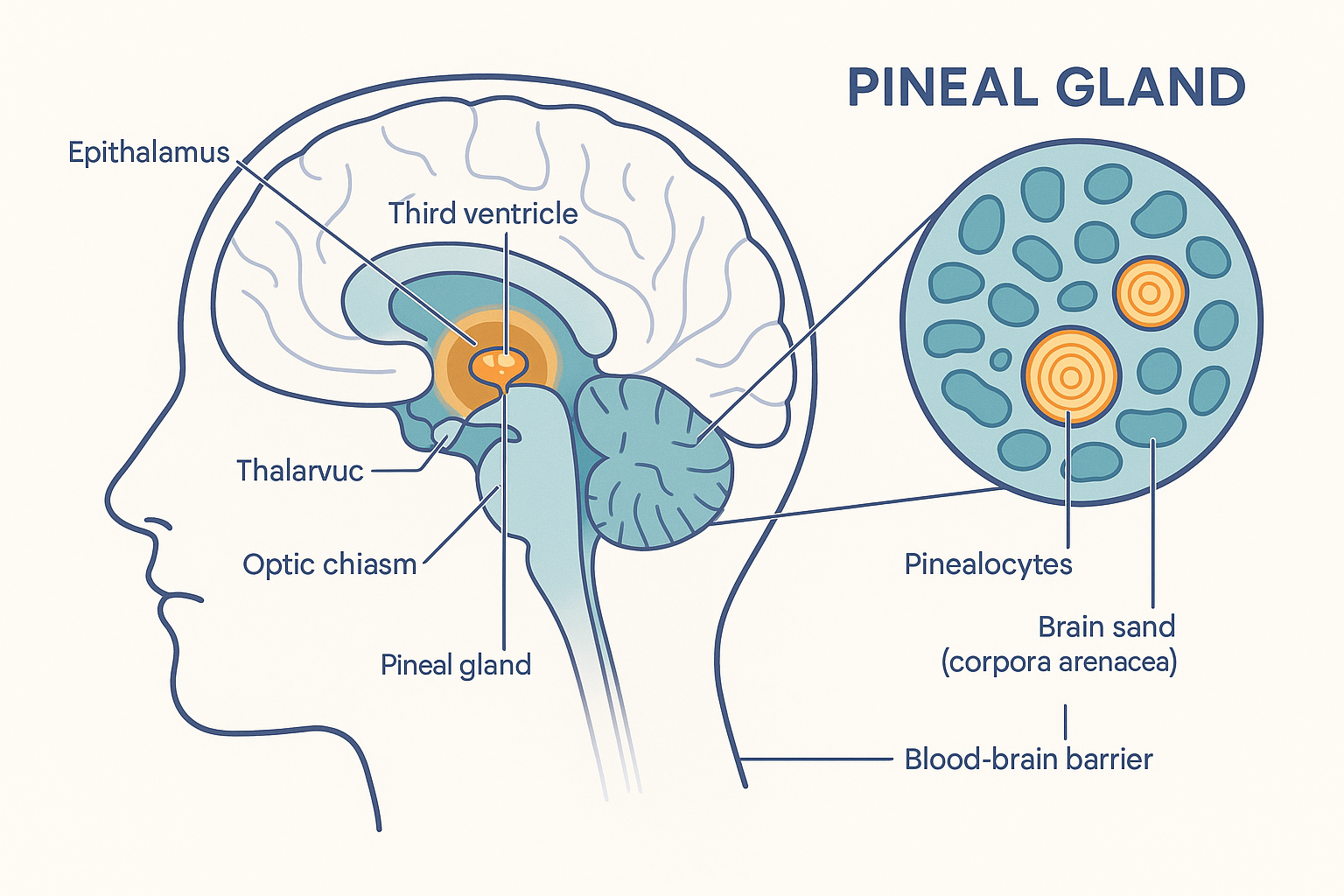 The pineal sits unpaired on the brain's midline at the back of the third ventricle, outside the blood-brain barrier — which lets it release melatonin straight into the blood but also exposes it to circulating calcium and fluoride that build up as "brain sand."
The core function: making melatonin from serotonin
The pineal's defining job is to synthesise melatonin (chemically, N-acetyl-5-methoxytryptamine), the hormone that signals "it is night" to the entire body. Melatonin is built in four steps from the dietary amino acid tryptophan, and — importantly — it travels down the same biochemical road as serotonin: serotonin is an intermediate on the way to melatonin. This is why the pineal pathway is best understood as an extension of serotonin synthesis, capped by two pineal-specific enzymes.
The pathway, step by step:
- Tryptophan → 5-hydroxytryptophan (5-HTP). Tryptophan, taken up from the blood, is hydroxylated by the enzyme tryptophan hydroxylase. This requires the cofactor BH4 (tetrahydrobiopterin) — the same cofactor that the dopamine pathway needs at its rate-limiting step (from the cognition page) — plus iron and oxygen.
- 5-HTP → serotonin. The enzyme aromatic amino acid decarboxylase (the same enzyme that turns L-DOPA into dopamine) removes a carboxyl group to make serotonin (5-hydroxytryptamine, 5-HT). This step requires vitamin B6 in its active form, P5P (pyridoxal-5-phosphate) — the identical B6-dependent decarboxylation seen in the catecholamine pathway. So far this is ordinary serotonin synthesis; the pineal simply holds a large pool of serotonin during the day, ready for the next steps.
- Serotonin → N-acetylserotonin (NAS). Here is the rate-limiting, light-controlled step — the whole system's master switch. The enzyme AANAT (arylalkylamine N-acetyltransferase), often called the "timezyme," attaches an acetyl group to serotonin. AANAT activity swings dramatically between day and night — by as much as 10- to 100-fold — and this oscillation is what makes melatonin a rhythmic, night-time hormone. When AANAT is switched off (by light, via the circuit below), melatonin production collapses regardless of how much serotonin is sitting in the gland.
- N-acetylserotonin → melatonin. The final enzyme, ASMT (acetylserotonin O-methyltransferase) — historically named HIOMT (hydroxyindole-O-methyltransferase) — adds a methyl group, producing melatonin. (The methyl group comes from SAM-e, the body's universal methyl donor, linking melatonin output loosely to methylation/folate status.) Unlike AANAT, ASMT is present at a fairly steady level throughout the day, so it does not gate the rhythm — it simply finishes whatever AANAT starts.
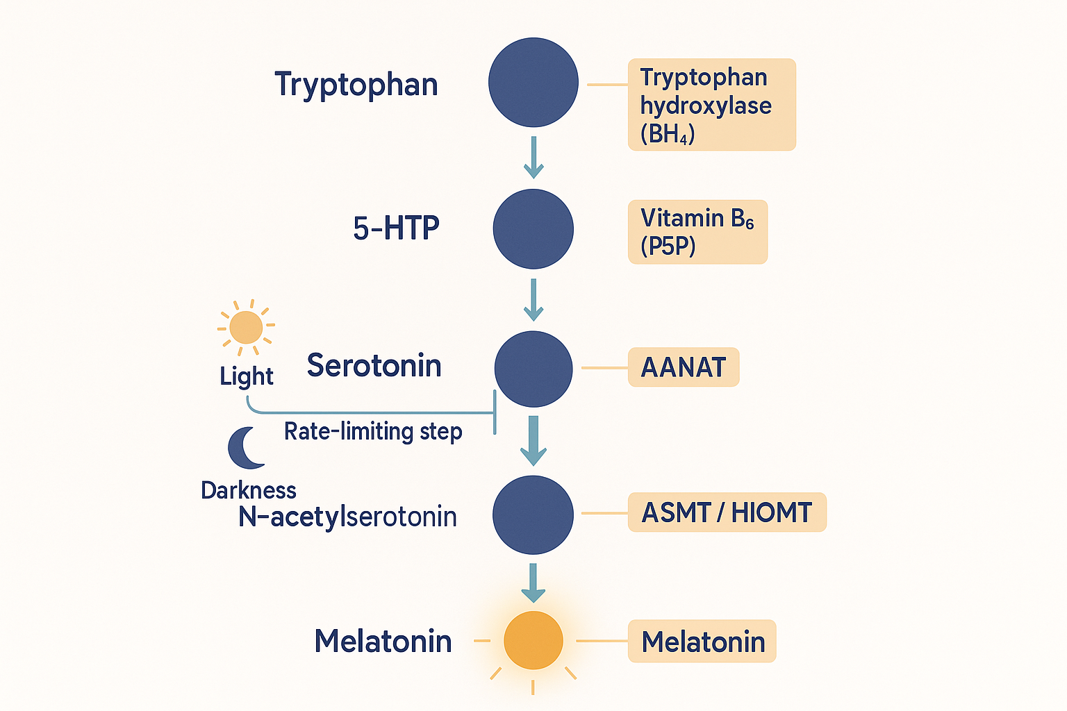 Melatonin is serotonin synthesis carried two steps further. The gatekeeper is AANAT — switched on in darkness and off by light — which is exactly why melatonin is a hormone of the night.
Why melatonin is made at night is now answerable precisely. The bottleneck enzyme, AANAT, is driven by a signal (noradrenaline) that the gland only receives in darkness, and is actively switched off when light hits the eye. So the gland is not "deciding" to make melatonin at night — it is being commanded to, by an external timing signal wired in from the eye. To see how that command arrives, we have to follow the circuit.
The wiring: from a photon to a hormone
The pineal in mammals is blind. It does not sense light itself; it is told about light by a chain of relays that starts in the retina and ends with a nerve squirting noradrenaline onto the gland. This is the part most worth understanding, because it explains the entire behaviour of the system. Trace it in order:
- The retina detects "day" with a special cell. Beyond the familiar rods and cones (which handle vision), the retina contains a small population of intrinsically photosensitive retinal ganglion cells (ipRGCs). These cells are not for seeing — they are for light measurement. They contain a pigment called melanopsin that is most sensitive to blue light (around 480 nm), the dominant wavelength of daylight (and, unfortunately, of screens and LED lighting). When blue light strikes them, ipRGCs fire. This is the body's true "is it daytime?" sensor, and it works even in some blind people who have lost all rod and cone vision.
- The signal travels to the master clock via a dedicated tract. The ipRGCs send their axons down a private pathway — the retinohypothalamic tract (RHT) — that branches off the optic nerve and goes not to the visual cortex but to a small cluster of neurons in the hypothalamus: the suprachiasmatic nucleus (SCN).
- The SCN is the master clock. The SCN (literally "above the optic chiasm," where the two optic nerves cross) is the body's central pacemaker — about 20,000 neurons that keep a self-sustaining ~24-hour rhythm and synchronise the whole body to the day/night cycle. Left in total darkness it keeps ticking on its own near-24-hour beat; the light signal from the retina is what resets it daily to exactly 24 hours and pins it to the real world (this resetting is entrainment). The SCN is the conductor; the pineal, as we will see, is one of the instruments it commands.
- The clock signals down into the spinal cord. From the SCN the timing signal passes to the paraventricular nucleus (PVN) of the hypothalamus, then descends through the brainstem into the spinal cord, to a column of sympathetic (fight-or-flight) neurons in the upper chest region — recall from the autonomic nervous system page that sympathetic nerves exit the cord in the thoracic segments.
- The signal loops up to a ganglion in the neck. Those spinal neurons connect to the superior cervical ganglion (SCG), a sympathetic relay station high in the neck. Postganglionic fibres from the SCG then travel back up into the skull and reach the pineal gland.
- Noradrenaline lands on the gland. At night, these sympathetic fibres release noradrenaline (norepinephrine) onto the pinealocytes. Noradrenaline binds β1-adrenergic receptors (with an α1 assist) on the cell surface, raising the second messenger cAMP, which activates PKA (protein kinase A), which switches on the transcription factor CREB — and this drives the production and stabilisation of AANAT, the rate-limiting enzyme. AANAT switches on, serotonin is converted, and melatonin pours out.
The crucial inversion to hold onto: light is inhibitory to melatonin. During the day, light keeps the ipRGCs firing, the SCN registers "daytime," and the sympathetic drive to the pineal is held off — so AANAT stays low and melatonin is negligible. At night, with no light, that brake is released, the SCN permits the sympathetic outflow, noradrenaline flows, AANAT rises, and melatonin climbs. And because the pathway runs through the light sensor in real time, a burst of light at night (a bright bathroom, a phone screen) signals "daytime" to the SCN and shuts AANAT off within minutes, collapsing melatonin. This single fact is the mechanistic basis for nearly all "light hygiene" advice: blue light at night is not vaguely "stimulating," it is directly switching off the enzyme that makes your sleep hormone.
flowchart TD
LIGHT["Blue light (~480 nm)<br/>daylight / screens"] --> IPRGC["ipRGCs in retina<br/>(melanopsin sensor)"]
IPRGC -->|retinohypothalamic tract| SCN["SCN — master clock<br/>(hypothalamus)"]
SCN --> PVN["Paraventricular nucleus"]
PVN --> CORD["Sympathetic neurons<br/>(thoracic spinal cord)"]
CORD --> SCG["Superior cervical ganglion<br/>(neck)"]
SCG -->|noradrenaline at night| PINE["Pinealocytes<br/>β1 receptor → cAMP → PKA → CREB → AANAT"]
PINE --> MEL["Melatonin released"]
LIGHT -.->|"light = STOP signal:<br/>shuts off AANAT in minutes"| PINE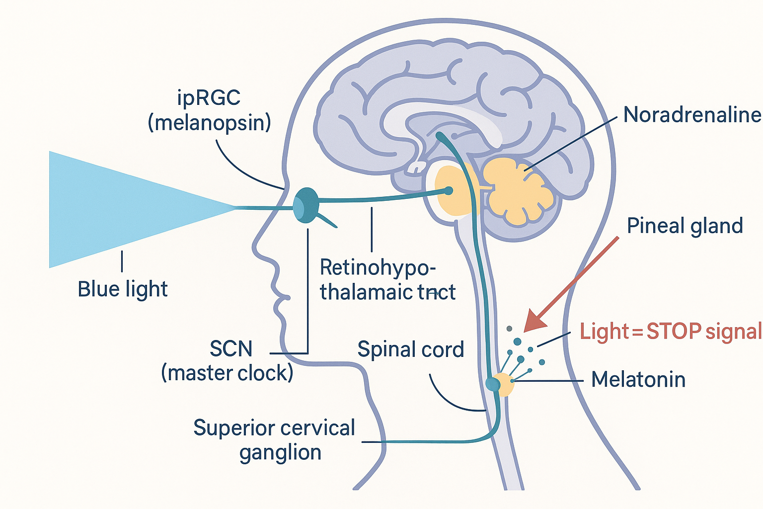 From photon to hormone: the eye measures light, the SCN keeps time, and a long sympathetic loop through the spinal cord and neck delivers the "make melatonin" command to the pineal at night — a command that light instantly cancels.
The molecular clock inside the SCN
How does the SCN "keep time" at all? Inside its neurons (and, it turns out, inside nearly every cell in the body) runs a molecular clock: a self-regulating genetic feedback loop that takes about 24 hours to complete one cycle. The mechanism is a transcription–translation negative feedback loop, and it is worth knowing because it is the actual gear-train of biological time:
- Two proteins, CLOCK and BMAL1, pair up and act as transcription factors — they switch on the genes named Period (PER) and Cryptochrome (CRY).
- The PER and CRY proteins are produced and accumulate in the cytoplasm through the day.
- After a delay, PER and CRY pair up, move into the nucleus, and block CLOCK/BMAL1 — shutting off their own genes. Production stops.
- The existing PER and CRY are then gradually degraded; with the brake gone, CLOCK/BMAL1 become active again and the cycle restarts. One full loop takes ~24 hours.
This loop is the cellular oscillator. The SCN's special role is that its thousands of neurons keep their loops synchronised with each other and reset by light — making the SCN the master pacemaker that imposes its rhythm on the millions of independent "peripheral clocks" elsewhere in the body (in the liver, gut, and so on). Melatonin is the SCN's principal hormonal output — the chemical broadcast it uses to tell the rest of the body what time the central clock thinks it is. In that sense the pineal is "the hormonal hand of the clock": the SCN is the movement, melatonin is the hand everyone can read.
flowchart LR
    CB["CLOCK + BMAL1<br/>(activators)"] -->|"switch ON genes"| PC["PER + CRY<br/>proteins made"]
    PC -->|"accumulate, enter nucleus"| INH["PER/CRY block CLOCK/BMAL1"]
    INH -->|"own genes shut off"| DEG["PER/CRY degrade over hours"]
    DEG -->|"brake released → restart"| CB
The ~24-hour molecular clock: CLOCK/BMAL1 turn on PER and CRY, which build up, feed back to switch their own genes off, then degrade so the cycle can start again. Light, via the SCN, keeps this loop set to the real day.
What melatonin actually does
Once released, melatonin is lipophilic (fat-soluble) and amphiphilic — it dissolves in both fat and water — so it spreads freely across every membrane and barrier into every tissue, cell, and organelle in the body. It has two broad classes of action: a receptor-mediated timing signal, and a direct chemical antioxidant effect that needs no receptor at all.
1. Setting the clock: the chronobiotic signal
Melatonin acts on two dedicated receptors, MT1 and MT2 — G-protein-coupled surface receptors (the fast, water-soluble-style receptors from the hormones page) — found densely on the SCN itself and throughout the body. Through them melatonin does several things:
- It feeds back onto the SCN to reinforce "night." Melatonin tells the master clock that darkness has arrived, helping consolidate the night-time state — the clock drives melatonin, and melatonin in turn steadies the clock.
- It promotes sleep onset. Rising evening melatonin "opens the sleep gate" — it does not sedate you the way a sleeping pill does (it is not a knock-out drug); it lowers the alerting drive and shifts the body toward sleep readiness. This is why exogenous melatonin works best as a timing cue at a modest dose, not a sedative at a large one.
- It is a chronobiotic — it shifts the clock's phase. This is melatonin's most powerful and underrated property. When you take it determines which way the clock moves (the "phase-response curve"): melatonin in the evening advances the clock (shifts everything earlier), while melatonin in the morning delays it (shifts later). This is precisely why melatonin is used — carefully timed — to treat jet lag and shift-work disruption: it is a lever on the clock's phase, not merely a sleep aid.
- It lowers core body temperature. Melatonin nudges blood flow toward the skin (distal vasodilation), shedding heat and dropping core temperature — itself a physiological signal for sleep. The natural night-time dip in body temperature is partly melatonin's doing.
2. The hidden job: a potent mitochondrial antioxidant
Less famous but increasingly seen as central: melatonin is one of the most effective antioxidants the body has — a molecule that neutralises reactive oxygen species (ROS), the destructive oxygen by-products of metabolism (recall from the cellular energy page that the electron transport chain inevitably leaks ROS). Several features make it unusually good at this:
- It is a direct free-radical scavenger. Melatonin chemically neutralises some of the most damaging radicals — the hydroxyl radical and peroxynitrite — directly, without needing a receptor.
- It is a "cascade" scavenger. When melatonin neutralises a radical, its breakdown products (AFMK and AMK) are also antioxidants, which neutralise further radicals in turn. One melatonin molecule can therefore mop up several radicals down a chain — unusual and efficient.
- It concentrates in mitochondria. Because it is lipophilic and amphiphilic, melatonin accumulates inside cells and especially in mitochondria — the exact site where ROS are generated. It protects the organelle at the source of the damage. (There is good evidence mitochondria can even synthesise their own melatonin locally, hinting this antioxidant role may be melatonin's original, most ancient function — older than its timekeeping job.)
- It boosts the body's own antioxidant enzymes. Beyond scavenging directly, melatonin upregulates protective enzymes such as superoxide dismutase and glutathione peroxidase, raising the cell's overall antioxidant capacity.
This dual identity — timing hormone by receptor, antioxidant by chemistry — is why the age-related fall in melatonin is viewed as more than just "worse sleep": it is also the loss of a nightly, body-wide, mitochondria-targeted antioxidant sweep.
The daily rhythm — and its decline with age
Putting the timing together: melatonin is essentially undetectable during the day, begins to rise a couple of hours before habitual sleep (the "dim-light melatonin onset," the body's internal dusk), peaks in the middle of the night (around 2–4 a.m.), and falls back toward zero by morning as light returns. That nightly pulse is the body's master timing signal — every tissue reads it.
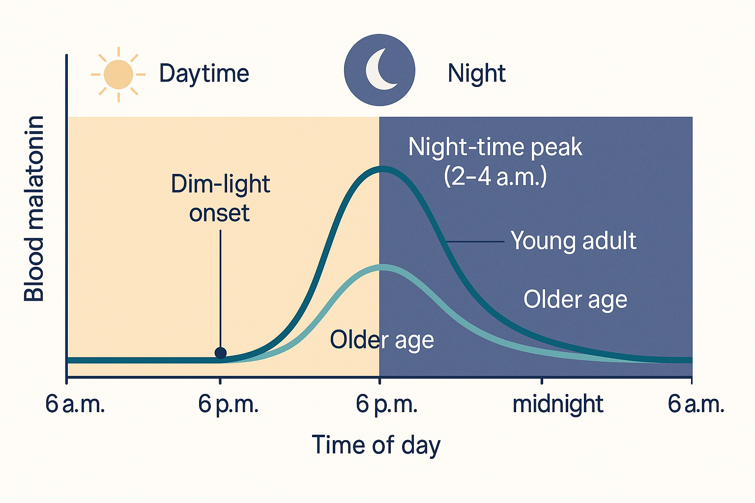 Melatonin traces a single nightly pulse — undetectable by day, peaking around 2–4 a.m. — and the amplitude of that peak shrinks markedly with age (faded curve), one of the clearest endocrine signatures of the ageing pineal.
The amplitude of this pulse falls steadily with age. Night-time melatonin output is highest in young children, declines through adolescence and adulthood, and in many elderly people the nocturnal peak is drastically blunted or nearly absent. Estimates vary, but the secretory capacity drops on the order of ~10–15% per decade from mid-life. Several things drive the decline: loss of secretory pinealocytes, reduced β-adrenergic receptor density (the gland hears the noradrenaline signal less well), gliosis, and — the part everyone asks about — calcification.
Calcification: the "brain sand"
If you look at a brain CT scan of almost any middle-aged adult, you will often see a small bright fleck on the midline: a calcified pineal gland. The gland accumulates concretions called corpora arenacea — literally "sandy bodies," and colloquially "brain sand." These are microscopic stones built up in concentric layers, made chiefly of hydroxyapatite (the same calcium-phosphate mineral as bone and tooth enamel) together with fluorapatite — hydroxyapatite in which fluoride has substituted into the crystal — plus an organic matrix and calcium/magnesium salts.
The key facts, calibrated to the evidence:
- It is extremely common and increases with age. Calcification appears as early as the second decade of life and becomes more prevalent and more extensive with each passing decade. Pooled across imaging studies, roughly 60% of adults show pineal calcification (estimates commonly run 40–70% depending on age and method). It is, in short, a near-universal feature of the ageing pineal — visible on CT, MRI, and even plain skull X-ray, where radiologists have long used the calcified pineal as a convenient midline marker (a shift of the calcified pineal off-centre signals a space-occupying lesion).
- It correlates with reduced melatonin. More calcification tends to track with lower melatonin output, and the degree of calcification rises alongside the age-related decline in melatonin. This makes biological sense: the calcified portion of the gland is non-secretory tissue. But correlation is the honest word — calcification and melatonin decline both advance with age and with the loss of functional pinealocytes, so it is not fully settled how much the calcification causes the melatonin fall versus the two being parallel symptoms of an ageing gland.
- Why the pineal in particular? Recall the gland sits outside the blood-brain barrier with a torrential blood supply — so it is bathed in circulating calcium and minerals more than most brain tissue, which plausibly predisposes it to mineral deposition. Recent work also implicates local inflammation in actively driving the calcification, rather than it being purely passive mineral settling.
The fluoride question — calibrated honestly
A persistent claim in wellness circles is that fluoride (from water, toothpaste) calcifies the pineal and suppresses melatonin. What is actually established, and what is not:
- What is real: The pineal does concentrate fluoride — strikingly so. Because it is outside the BBB and heavily perfused, the gland accumulates fluoride to among the highest concentrations of any soft tissue in the body, and that fluoride is incorporated into the calcified deposits as fluorapatite (confirmed by mineral analysis of human glands). So the gland genuinely is a fluoride sink, and fluoride genuinely is part of the brain sand's chemistry.
- What is suggestive but not proven: A frequently cited animal study (Luke, in gerbils) found that higher pineal fluoride was associated with lower melatonin output and earlier puberty — a plausible mechanistic hint that fluoride loading might blunt the gland. But this is limited animal data; robust human evidence that ordinary fluoride exposure causes meaningful melatonin suppression is lacking.
- The honest bottom line: Fluoride accumulates in the pineal and is part of its calcification — that much is solid. The leap from there to "fluoride is shutting down your pineal and you must 'decalcify' it" is not supported by strong human evidence, and the various "decalcification" protocols sold online have no demonstrated ability to remove established calcification or restore melatonin. Pineal calcification is best understood as a real, near-universal, age-related, multifactorial process in which fluoride is one contributing player — not the cartoon villain, and not something a supplement dissolves away.
Longevity, sleep, and the pineal peptides — calibrated
Because melatonin is both the body's master timing signal and a nightly mitochondrial antioxidant, the argument that a well-functioning pineal rhythm matters for healthy ageing is mechanistically reasonable. A robust nocturnal melatonin pulse supports consolidated sleep (during which the brain's glymphatic clearance and the body's repair programmes run), reinforces the circadian organisation that keeps peripheral clocks aligned, and delivers an antioxidant sweep to the mitochondria each night. The flip side — the blunted, arrhythmic melatonin of old age, shift work, and chronic light-at-night — is associated with poorer sleep, metabolic disruption, and is an active area of research in neurodegeneration (where circadian and melatonin disturbances appear early in conditions like Alzheimer's, though whether as cause or consequence is unresolved).
This is the context for the Khavinson pineal peptides, which the biohacking community is most interested in:
- Epitalon (Epithalon, AEDG) — a synthetic tetrapeptide modelled on a pineal extract, whose headline claims are activation of telomerase (the enzyme that maintains chromosome end-caps) and restoration of the pineal's own melatonin output — that is, coaxing the ageing gland to secrete more melatonin again, rather than supplying melatonin from outside.
- Pinealon (EDR) — a related pineal-derived tripeptide pitched more at acute neuroprotection and cognition than at longevity.
The mechanisms, evidence, dosing, the all-important single-lab caveat (most data originate from Khavinson's own group), the 2025 independent replication of the telomere mechanism, and the honest hype-versus-evidence calibration are covered in full in the Pinealon & Epithalon deep dive — the central tie-in to this page. The short version for calibration: the idea that the pineal's decline is a meaningful node in ageing is plausible and mechanistically grounded; the specific claim that these peptides restore a youthful pineal in humans is promising but under-evidenced, and should be held with appropriate scepticism. The most reliable lever on your pineal remains the boring one the wiring diagram demands: bright light by day, darkness at night.
Putting it all together
- The pineal gland is a tiny (~5–8 mm, ~100–150 mg), unpaired, midline endocrine gland in the epithalamus at the back of the third ventricle, built mostly of pinealocytes, sitting outside the blood-brain barrier with a huge blood supply — so it releases hormone straight to the blood but is also exposed to circulating minerals.
- Its job is to make melatonin, built in four steps from tryptophan down the serotonin pathway (tryptophan → 5-HTP → serotonin → N-acetylserotonin → melatonin), gated by the rate-limiting enzyme AANAT, which is switched on by darkness and off by light — which is why melatonin is a night-time hormone.
- The gland is blind and is told about light by a long circuit: blue light → melanopsin ipRGCs in the retina → SCN (the master clock) → paraventricular nucleus → thoracic spinal cord → superior cervical ganglion → noradrenaline onto pinealocytes (β1 → cAMP → PKA → CREB → AANAT). Light is inhibitory — a flash at night shuts melatonin off within minutes.
- The SCN keeps time with a ~24-hour CLOCK/BMAL1 ↔ PER/CRY genetic feedback loop, reset daily by light; melatonin is the SCN's hormonal output — "the hand of the clock."
- Melatonin acts two ways: as a chronobiotic timing signal via MT1/MT2 receptors (sleep onset, phase-shifting that treats jet lag, lowering core temperature), and as a potent, mitochondria-targeting, cascade antioxidant that needs no receptor.
- Output falls with age — the nocturnal peak is blunted or lost in many elderly people — driven by pinealocyte loss, fewer β-receptors, and calcification.
- Calcification ("brain sand," corpora arenacea) is near-universal (~60% of adults, from the second decade up), made of hydroxyapatite/fluorapatite, correlates with lower melatonin, and is visible on imaging. The fluoride angle is real but overstated: the gland concentrates fluoride and incorporates it into the deposits, but strong human evidence that ordinary fluoride suppresses melatonin — or that "decalcification" works — is lacking.
- The longevity/pineal-peptide angle (Epitalon's melatonin/telomerase claims, Pinealon's neuroprotection) is mechanistically plausible but under-evidenced — see the deep dive — and the proven lever on pineal health remains light by day, darkness by night.
The unifying idea: the pineal gland is a transducer. It takes an environmental signal — the presence or absence of light, measured by the eye and timed by the SCN — and converts it into a chemical one, melatonin, broadcast to every cell. It is where the outside world's clock becomes the body's clock. Understand the circuit, and the whole of sleep timing, jet lag, light hygiene, the antioxidant role, and the ageing of the gland fall out of one elegant chain that runs from a photon to a hormone.
Related Compounds & Deep Dives
The pineal hormone and its pathway
- Melatonin — the pineal's output hormone; the chronobiotic timing signal and mitochondrial antioxidant described throughout this page.
- Tryptophan / L-tryptophan — the dietary amino acid that is the raw material for the entire serotonin → melatonin pathway.
- P5P (vitamin B6) — the active-B6 cofactor for the decarboxylation step that makes serotonin en route to melatonin.
Pineal-targeting peptides (the central tie-in)
- Pinealon & Epithalon deep dive — the Khavinson pineal bioregulators; Epitalon's melatonin-restoration and telomerase claims, Pinealon's neuroprotection, with full evidence calibration.
- Epitalon — the pineal-derived tetrapeptide claimed to restore endogenous melatonin and activate telomerase.
- Pinealon — the pineal-derived tripeptide pitched at acute neuroprotection and cognition.
Light, rhythm, and supporting inputs
- Circadian rhythm — the broader timing system the pineal serves.
- Sunlight / light therapy — bright daytime light is the primary entraining signal for the SCN and the strongest lever on pineal rhythm.
- Magnesium — a cofactor and calming mineral supporting sleep architecture (see the Magnesium deep dive).
Related foundations
- Hormones & the Endocrine System — the pineal as one of the endocrine glands, and the receptor logic (MT1/MT2 as surface receptors) that melatonin obeys.
- Autonomic Nervous System — the sympathetic wiring (noradrenaline, the superior cervical ganglion) that actually drives the gland at night.
- Neuroscience of Cognition — the shared serotonin/catecholamine biochemistry, BH4 and B6 cofactors, and the SCN's place in the wider brain.
- Cellular Energy — the mitochondrial ROS that melatonin's antioxidant role exists to neutralise.