Glycation, AGEs & Ageing
The slow caramelisation of you. Sugar does not just feed you — it sticks to you, chemically and permanently, browning your proteins the way heat browns a steak. This page is about that reaction: how a reducing sugar latches onto a protein with no enzyme telling it to, how the bond hardens over weeks into something the body can never undo, and why the gradual accumulation of these sugar-damaged proteins is one of the two great non-enzymatic engines of ageing — the sugar-driven twin of oxidative stress.
In 1912 a young French physician-chemist, Louis-Camille Maillard, described what happens when you gently heat a mixture of a sugar and an amino acid: it turns brown, develops aroma, and forms a cascade of new compounds. He was looking for clues about how the body builds proteins; what he had actually found was the chemistry of the crust on bread, the sear on meat, the colour of roasted coffee and beer. For half a century the Maillard reaction belonged to food science. Then, in the 1960s and 70s, came the unsettling realisation that the same reaction runs inside the living body — slowly, at 37 °C, with blood glucose as the sugar and your own proteins as the amino partner. The pivotal clue was haemoglobin. In 1968 the biochemist Samuel Rahbar noticed an unusual "fast" haemoglobin band that was markedly elevated in people with diabetes. By the late 1970s and early 1980s this band — glycated haemoglobin, or HbA1c — had been identified as haemoglobin with a glucose molecule stuck onto it, and was adopted as the standard measure of long-term blood-sugar control. The headline diabetes test is, at bottom, a measurement of how much glycation is happening inside you.
The 1980s and 90s opened the third act. Researchers showed that early sugar-protein adducts mature, over weeks to years, into a heterogeneous family of stable end-products — Advanced Glycation End-products (AGEs) — that cross-link long-lived structural proteins and stiffen tissues. Then a receptor for them was discovered and cloned (1992): RAGE (Receptor for AGEs), which converts the presence of these damaged proteins into an active inflammatory signal. Today the field runs in three directions at once: anti-glycation chemistry (compounds that scavenge the reactive intermediates), AGE cross-link breakers (molecules that try to cut the hardened bridges already formed), and a large and genuinely contested dietary-AGE epidemiology asking how much the browning on your dinner actually contributes to the AGEs in your arteries. This page builds the whole story from first principles.
Why this page exists
The Systemic Metabolism page already owns the story of how the body handles glucose — how insulin partitions it, how the liver buffers it, why insulin resistance lets blood sugar drift up. This page is not about handling sugar. It is about the damage sugar does on contact — a chemical process that proceeds whether or not your metabolism is "working", simply because glucose is a reactive molecule and your proteins are sitting in it.
This matters because it reframes "high blood sugar" from an abstract number into a mechanism of physical harm. When a clinician says chronic hyperglycaemia "damages the vessels, the kidneys, the nerves, the retina, the lens", glycation is a large part of what they mean — literally. And it reframes ageing: alongside the oxygen-driven wear-and-tear of oxidative stress, glycation is the other major way your structural proteins degrade without any enzyme being involved, slowly converting supple, functional tissue into stiff, cross-linked, dysfunctional tissue. The two processes are not independent — they feed each other, in a vicious cycle called glycoxidation that this page will make explicit.
We start by drawing a hard line between two things that share a name and are constantly confused.
Glycation is not glycosylation — the crucial distinction
Two words, almost identical, opposite in meaning. Getting them straight is the foundation of everything that follows.
Glycosylation (with an s) is a controlled, enzymatic, functional process. The body deliberately attaches sugar chains to specific proteins and lipids using dedicated enzymes — glycosyltransferases — that place a particular sugar at a particular site for a particular purpose. Most proteins on the outside of your cells and most secreted proteins are glycosylated this way; the sugar coat is part of the protein's design, used for folding, recognition, signalling, and stability. It is a feature. The cell builds it on purpose, the way a factory chrome-plates a part.
Glycation (no s) is the opposite in every respect: uncontrolled, non-enzymatic, and damaging. No enzyme is involved. No site is chosen. A reducing sugar simply collides with an amino group on a protein and, by ordinary chemistry, sticks. It is not a feature; it is corrosion. The cell does not want it, cannot direct it, and largely cannot reverse it. Where glycosylation is chrome-plating, glycation is rust.
The single distinction to hold onto: glycosylation is the cell's deliberate decoration of proteins with sugar; glycation is the accidental, damaging caramelisation of proteins by loose sugar. This page is entirely about the second one. Everywhere below, "glycation" means the rust, never the chrome.
What does "non-enzymatic" actually require? Just two ingredients in contact: a reducing sugar (a sugar with a reactive open-chain form bearing a free carbonyl group — glucose, fructose, galactose, ribose all qualify) and a free amino group on a protein, lipid, or even DNA. Amino groups are everywhere: the side-chain of every lysine residue, the guanidino group of every arginine, and the N-terminus of every protein chain all carry them. Put a reactive sugar next to these groups in a warm, watery environment — which is to say, put sugar in a body — and the reaction proceeds spontaneously. It needs no catalyst because it is simply thermodynamically downhill; the only variables are how much sugar, how reactive that sugar is, and how long the protein sits there exposed. That last variable — time — is why long-lived proteins are the principal victims, a point we return to.
The chemistry: Schiff base → Amadori product → AGE
The reaction is not a single step but a slow, staged maturation. Each stage is more stable and more permanent than the last. Understanding the three stages explains nearly everything about why glycation is dangerous and where it can be intercepted.
Stage 1 — the Schiff base (fast, freely reversible, minutes to hours). The open-chain form of glucose carries an aldehyde group (a carbon double-bonded to oxygen, with a hydrogen attached: –CHO). This carbon is electron-poor and electrophilic — it actively seeks electrons. The lone pair of electrons on a protein's amino group (–NH₂) attacks it, and the two condense, losing a molecule of water, to form a Schiff base (a carbon–nitrogen double bond, –CH=N–; also called an aldimine). This first adduct is unstable and fully reversible: it forms in minutes and falls apart again just as readily. Its concentration simply tracks the surrounding sugar concentration — high glucose, more Schiff base; sugar falls, the Schiff base dissolves back. At this stage nothing permanent has happened.
Stage 2 — the Amadori product (slow, stable, but still reversible; hours to weeks). Some Schiff bases, instead of falling apart, undergo an internal rearrangement — the Amadori rearrangement — into a more stable arrangement of atoms (a ketoamine). This is the pivotal step. The Amadori product is far more stable than the Schiff base; it does not readily fall apart when sugar levels drop, so it accumulates in proportion to the time-averaged sugar exposure. It is still, in principle, reversible and is not yet an AGE — but it is the long-lived early footprint of glycation, and it is exactly what HbA1c measures. HbA1c is the Amadori product of glucose on haemoglobin. Hold that thought; it is the most concrete example in the whole topic and we develop it fully below.
Stage 3 — Advanced Glycation End-products (slow, irreversible; weeks to years). Over weeks and months, Amadori products undergo a thicket of further reactions — oxidation, dehydration, fragmentation, rearrangement, and condensation — to form AGEs: a chemically diverse family of stable, often coloured, often fluorescent compounds. This stage is, for practical purposes, irreversible — the body has no general enzyme that reverses a mature AGE. Some AGEs are simple adducts sitting on a single residue, the best-known being carboxymethyl-lysine (CML), the most widely used AGE biomarker. Others are far more consequential: cross-links that covalently bridge two proteins together. Glucosepane (an arginine–lysine bridge) is the most abundant cross-link in aged human tissue; pentosidine is the most studied. A cross-link is the molecular event that converts a supple tissue into a stiff one, and it is where glycation does its structural damage.
flowchart LR
GLU["Reducing sugar<br/>(open-chain glucose, -CHO)"] --> SB
NH2["Protein amino group<br/>(lysine / arginine / N-terminus)"] --> SB
SB["Schiff base<br/>(minutes; freely reversible)"] -->|Amadori rearrangement| AM["Amadori product<br/>(weeks; stable, still reversible)<br/>= HbA1c on haemoglobin"]
AM -->|"oxidation, dehydration,<br/>fragmentation (weeks–years)"| AGE["AGE<br/>(irreversible)"]
AGE --> ADD["Adducts<br/>e.g. CML"]
AGE --> XL["Cross-links<br/>glucosepane, pentosidine<br/>= tissue stiffening"]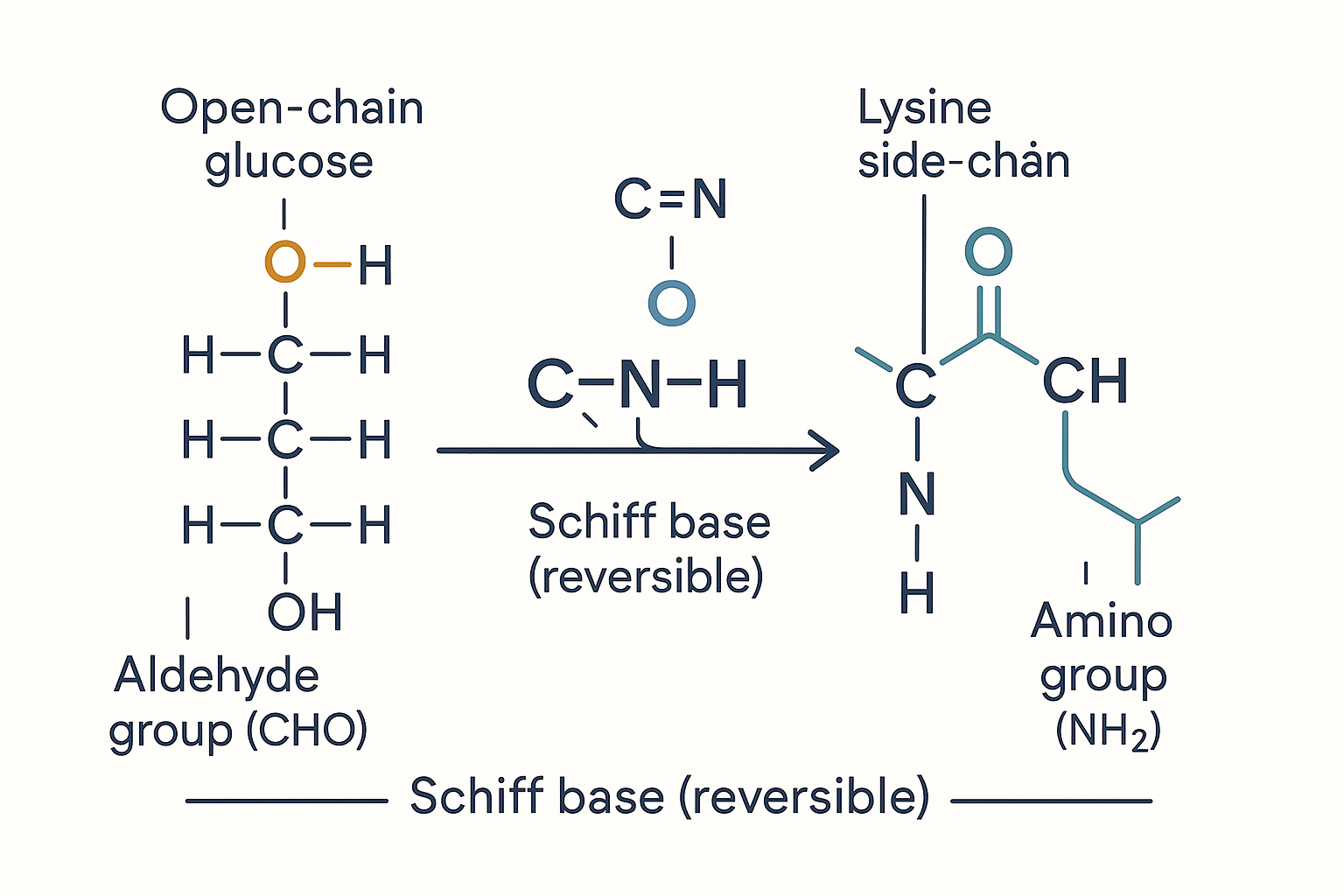 The molecular act of glycation: glucose's open-chain aldehyde group reacts with a protein's free amino group, condensing into a Schiff base and releasing water — the first, reversible step before the bond matures into a permanent AGE.
The dicarbonyl shortcut — the fast lane to AGEs
There is a faster, nastier route that bypasses the slow staged maturation. Glucose metabolism and the breakdown of Schiff bases and Amadori products throw off a class of small, ferociously reactive molecules called dicarbonyls — most importantly methylglyoxal (MGO), plus glyoxal and 3-deoxyglucosone. A dicarbonyl carries two carbonyl groups close together, which makes it dramatically more reactive than glucose itself — methylglyoxal glycates proteins on the order of thousands of times faster than glucose. These molecules are the most potent endogenous AGE precursors, hammering arginine and lysine residues directly and rapidly into AGEs (MGO-derived hydroimidazolone is among the commonest AGEs in the body).
Because dicarbonyls are so dangerous, the body invests in a dedicated defence: the glyoxalase system (glyoxalase 1 and 2, GLO1/GLO2), a pair of enzymes that, using glutathione as a cofactor, detoxify methylglyoxal into harmless lactate before it can do damage. This is a direct and important bridge to the oxidative stress page: the glyoxalase defence runs on glutathione, the same master antioxidant that defends against oxygen radicals. When glutathione is depleted — by oxidative stress, by ageing, by toxic load — the glyoxalase brake weakens and methylglyoxal-driven glycation accelerates. Sugar damage and oxygen damage share a defence budget, and draining one drains the other.
Why fructose is the worse sugar
Not all reducing sugars are equal. Fructose glycates far faster than glucose — roughly 7.5 to 10 times faster in head-to-head tests. The reason is structural: glycation can only happen via a sugar's open-chain reactive form, and glucose is overwhelmingly locked in a stable ring — only about 0.002% of glucose exists in the open-chain aldehyde form at any instant. Glucose is, in a sense, a deliberately sluggish sugar; its ring-favouring chemistry is part of why evolution chose it as the main blood sugar. Fructose holds a much larger fraction in its reactive open-chain form and carries a keto group that feeds readily into dicarbonyl formation, so molecule-for-molecule it is a far more aggressive glycating agent.
This deserves an honest calibration, because the popular framing over-reaches. Despite fructose's higher intrinsic reactivity, normal blood fructose is very low (the liver clears dietary fructose on first pass), so fructose contributes relatively little to glycation in the bloodstream compared with glucose, which is present at far higher concentration. Where fructose's reactivity bites hardest is inside cells and tissues that are exposed to high fructose loads or that generate fructose internally — notably the liver under a high-sugar/HFCS diet, and tissues that run the polyol pathway (which converts glucose to fructose internally; this is a major mechanism of diabetic damage in the lens, nerves, and kidneys, where local fructose and its dicarbonyl by-products glycate proteins aggressively). So the accurate statement is not "fructose in your blood is glycating you ten times faster than glucose" — it is "fructose is an intrinsically far more dangerous glycating sugar, and the tissues exposed to high fructose pay the price." This is the molecular core of why high-sugar and high-fructose-corn-syrup diets are implicated in accelerated tissue ageing.
Endogenous AGEs: the damage your own blood sugar does
"Endogenous" means generated inside the body, from your own circulating sugar — and this is the dominant, best-established source of biologically important AGEs. The driver is simple and quantitative: the higher and more sustained your blood glucose, the faster you glycate. Someone with chronic hyperglycaemia is bathing every exposed protein in more sugar for more hours of the day, and the glycation reaction, being non-enzymatic and concentration-driven, simply runs faster. This is why diabetes is, at the molecular level, the canonical AGE disease.
HbA1c: the diabetes test is a glycation measurement
This is the concept to anchor the whole page on, because it makes the abstract chemistry concrete and clinical. Haemoglobin is the oxygen-carrying protein packed inside red blood cells. It sits, for the entire ~120-day lifespan of the red cell, bathed directly in blood glucose — and glucose glycates it. Specifically, glucose forms an Amadori product on the N-terminal valine of the haemoglobin beta-chain. The result is HbA1c — glycated haemoglobin — and the percentage of a person's haemoglobin that is glycated is directly proportional to the average glucose concentration their blood has been carrying.
Because the red cell lives about three to four months and cannot repair its haemoglobin, HbA1c is a running average of blood sugar over the preceding ~8–12 weeks — immune to the day-to-day swings that a single glucose reading catches. A normal HbA1c is below about 5.7%; diabetes is diagnosed at 6.5% or above. Every one of those percentage points is a direct readout of how much sugar has chemically stuck to a protein inside you. The single most important blood test in diabetes — the number that predicts complications and guides treatment — is, mechanistically, a glycation assay. Internalise this and the rest of the page stops being abstract: HbA1c proves that glycation is happening in your body right now, continuously, at a rate set by your blood sugar.
The crucial extrapolation: haemoglobin is replaced every few months, so its glycation resets. But many structural proteins are not replaced for years or decades — and those are where glycation accumulates without limit.
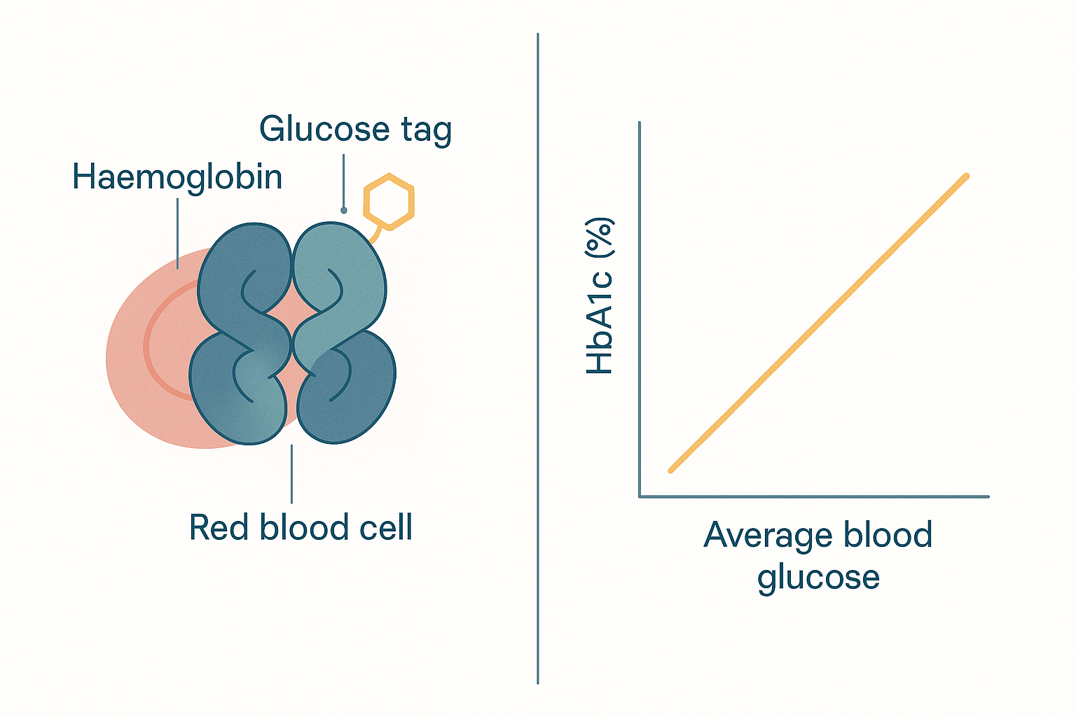 HbA1c is glycated haemoglobin: glucose forms a stable Amadori adduct on haemoglobin over the red cell's ~120-day life, so the percentage glycated tracks average blood glucose over ~2–3 months — the standard diabetes test is, at bottom, a measurement of glycation.
Collagen cross-linking: how glycation stiffens you
Here is where glycation becomes ageing you can feel. The body's long-lived structural proteins — above all collagen (the scaffold of skin, arteries, tendons, bone, and basement membranes) and elastin (the protein that lets tissues spring back) — turn over extremely slowly. Skin and arterial collagen can persist for a decade or more; some structural proteins last essentially a lifetime. The longer a protein lives, the longer it is exposed to glucose, and the more AGE cross-links accumulate on it. And cross-links do something mechanically specific: they covalently weld adjacent collagen fibres together, converting a tissue's organised, sliding, flexible fibre bundles into a rigid, fused mesh.
The consequences map directly onto the visible and measurable signatures of ageing:
- Arterial stiffening. Cross-linked arterial collagen loses its elastic give. Stiff arteries cannot buffer the pressure pulse from each heartbeat, which raises systolic blood pressure and pulse pressure and strains the heart. AGE-driven arterial stiffening is a major reason vascular age outpaces calendar age in diabetes, and a contributor to age-related hypertension generally.
- Skin wrinkling and loss of elasticity. Glycated, cross-linked dermal collagen and elastin become stiff and brittle; the skin loses its snap-back and resilience. This is the genuine mechanism behind the popular "sugar sag" framing of skin ageing — and unlike most cosmetic claims, it rests on real chemistry (more on the calibration below).
- Tendon, ligament, and joint stiffening. The reduced suppleness and increased brittleness of aged connective tissue is partly AGE cross-linking; it contributes to stiffness and to the reduced resilience of cartilage in osteoarthritis.
- Cataracts — the cleanest example of all. The proteins of the eye's lens, the crystallins, are laid down before birth and never replaced — they must last your entire life. That makes them the ultimate sitting target. Over decades they accumulate glycation and oxidation, cross-link, aggregate, and scatter light — and the lens clouds. This is a cataract, and it is accelerated dramatically by diabetes precisely because higher sugar means faster glycation of proteins that can never be renewed. The lens is glycation's purest demonstration: a tissue that cannot reset, browning slowly over a lifetime exactly like Maillard's heated sugar-protein mixture, only at body temperature over decades.
The classic diabetic complications are this same process concentrated by high sugar: nephropathy (kidney damage — glycation of the glomerular basement membrane), retinopathy (damage to the retinal microvasculature), and neuropathy (nerve damage). Diabetes is, in large part, accelerated AGE disease — which is why it doubles as a model of accelerated ageing.
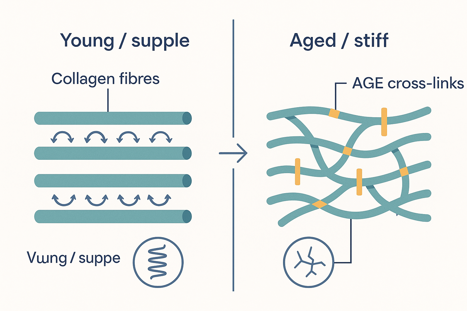 The visual essence of why glycation ages you: AGE cross-links covalently weld adjacent collagen fibres together, converting supple, elastic, sliding tissue (left) into a stiff, brittle, fused mesh (right) — the molecular basis of arterial stiffening, skin wrinkling, joint stiffness, and cataract.
Dietary AGEs: the cooking angle
So far every AGE has been made inside you from your own blood sugar. But AGEs also arrive pre-formed in food, because cooking is the Maillard reaction run deliberately and at high temperature. This is the exogenous (from outside) source, and it is where the science is genuinely less settled — so it deserves careful, calibrated treatment rather than the alarmism it often attracts.
Cooking method is the dominant variable — far more than the food itself. The Maillard browning reaction needs heat and dryness; it accelerates sharply with temperature and is suppressed by water (which caps the temperature at 100 °C and dilutes the reactants). The practical hierarchy is large and reliable:
- Low-AGE methods — wet and gentle: steaming, boiling, poaching, stewing, slow-cooking. Water keeps the temperature down and browning minimal.
- High-AGE methods — dry and hot: grilling, frying, roasting, searing, broiling, barbecuing. The browned, crusted, charred surfaces are exactly where AGEs are concentrated. The same chicken breast can carry several-fold more AGEs grilled than poached.
Beyond AGEs proper, high-heat dry cooking of starchy foods generates acrylamide — a separate browning by-product formed from the amino acid asparagine reacting with reducing sugars above ~120 °C, found in chips/fries, crisps, toast crusts, and roasted coffee. Acrylamide is a probable carcinogen and a distinct concern from AGEs, but it travels in the same company: it is another reason the charred and deep-browned end of the cooking spectrum is the one to moderate.
Now the honest calibration, because this is where popular writing overreaches. How much do dietary AGEs actually matter? The evidence is real but unsettled. Several things are reasonably established: only a fraction of ingested AGEs is absorbed — roughly 10–30% crosses the gut, and only about a third of that is excreted in urine, with the rest distributing into tissue; many food AGEs are large molecules that pass to the colon and interact with gut bacteria instead; and controlled feeding studies have shown that high-AGE diets raise circulating AGE markers and inflammatory markers in humans, while AGE-restricted diets lower them. That is genuine signal. But the relative weight of diet versus endogenous production is actively debated: some researchers argue dietary AGEs are a major contributor to the body's AGE pool, others that endogenous formation from your own blood sugar dominates and dietary AGEs are a smaller, modifiable add-on. The methodological problems are real (AGE measurement is not standardised; food AGE databases are imperfect).
The defensible synthesis: endogenous AGEs driven by your own blood sugar are the larger and better-established lever; dietary AGEs are a real, plausibly meaningful, and easily modifiable secondary contributor. Favouring gentle wet cooking over charring is a sensible, low-cost hedge — but it is the junior intervention. Lowering chronic blood glucose is the senior one. Anyone selling dietary-AGE avoidance as the master key to ageing has the hierarchy backwards.
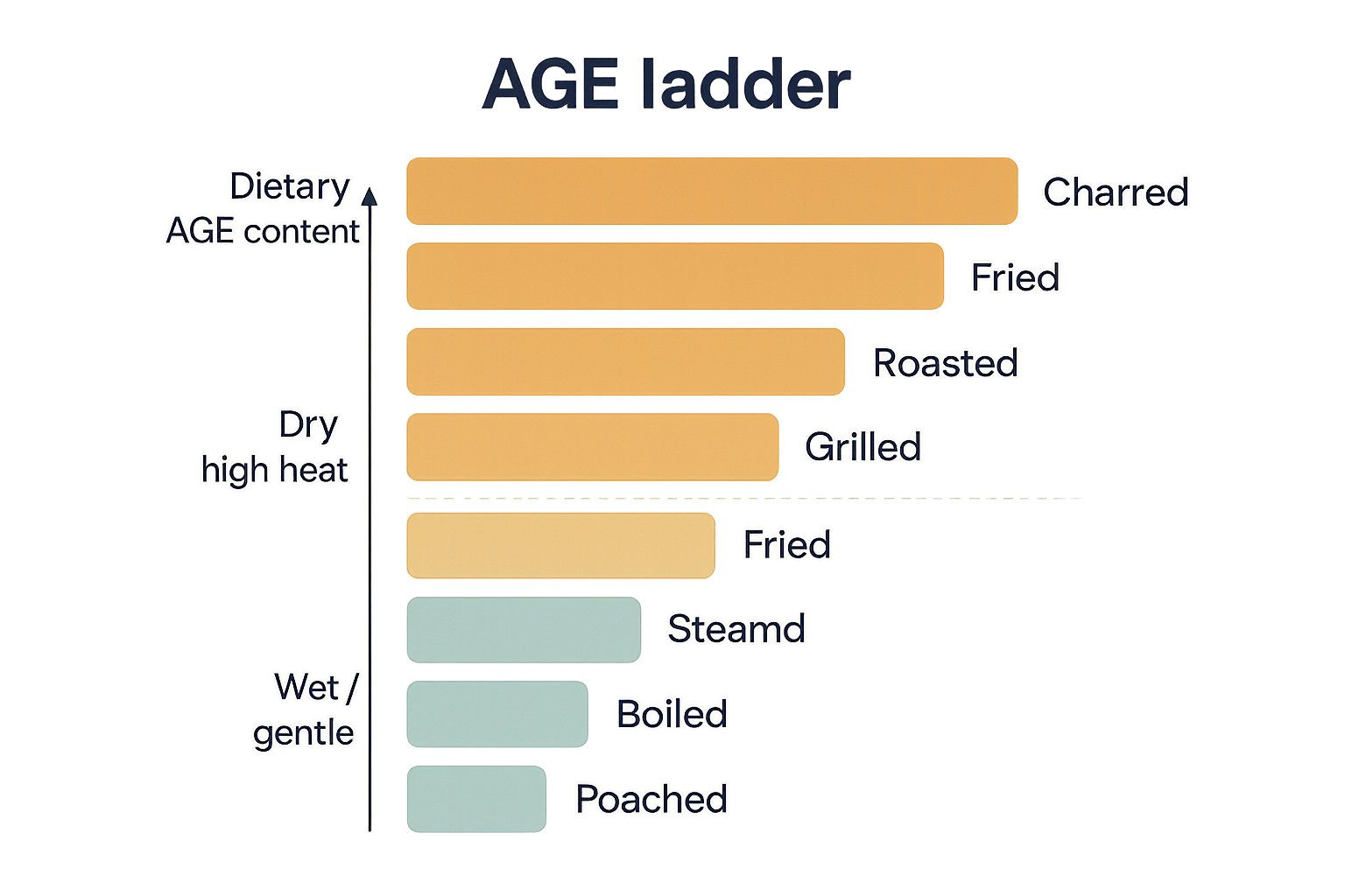 Cooking method sets dietary AGE load far more than the food itself: wet, gentle methods (steaming, boiling, poaching) generate little browning; dry, high-heat methods (roasting, grilling, frying, charring) generate many times more — a real but secondary lever next to controlling blood glucose.
RAGE and the glycoxidation loop
AGEs would be bad enough if they were merely inert structural damage — stiff collagen, clouded lenses. But they are worse than inert: the body has a receptor that actively detects them and responds with inflammation. This is RAGE — the Receptor for Advanced Glycation End-products — a surface receptor on endothelial cells (the vessel lining), immune cells, and many others.
When an AGE binds RAGE, the receptor triggers an intracellular signal that activates NF-κB — the master inflammatory switch introduced in full on the inflammation page. Activated NF-κB enters the nucleus and turns on the genes for inflammatory cytokines (TNF-α, IL-6 and others) and for enzymes that generate reactive oxygen species. In other words, AGEs do not just sit there as damage — they are read as a danger signal and converted into active, low-grade, chronic inflammation. This is a direct mechanistic bridge to the inflammation page: AGEs are one of the persistent triggers that keep the inflammatory fire smouldering, and RAGE is the sensor that lights it.
Two features make this genuinely vicious:
-
RAGE upregulates itself. Activating NF-κB increases the expression of RAGE itself (its gene is an NF-κB target). So AGE binding makes more receptor, which catches more AGEs, which makes more inflammation — a feed-forward amplifier rather than a self-limiting response. Unlike a well-behaved signalling system that desensitises, RAGE turns the volume up on continued exposure.
-
Glycoxidation — the loop with oxidative stress. This is the central reason glycation and oxidation are twins rather than rivals. RAGE activation generates reactive oxygen species (partly by switching on the enzyme NADPH oxidase and by stressing mitochondria). Those ROS are exactly the agents of oxidative stress — and oxidative conditions, in turn, accelerate the formation of AGEs (many AGE-forming steps, including CML formation and dicarbonyl generation, are oxidation reactions) and deplete glutathione, which weakens the glyoxalase defence against methylglyoxal. So: sugar damage triggers oxygen damage triggers more sugar damage. Each amplifies the other in a self-reinforcing spiral. The umbrella term for AGE formation occurring through oxidative chemistry is glycoxidation, and the loop is why the two great non-enzymatic ageing processes are best understood together rather than apart.
flowchart TD
    AGE["AGEs<br/>(circulating + tissue-bound)"] --> RAGE["RAGE receptor<br/>(endothelium, immune cells)"]
    RAGE --> NFKB["NF-κB activated<br/>(master inflammatory switch)"]
    NFKB --> CYTO["Cytokines<br/>TNF-α, IL-6"]
    NFKB --> ROS["Reactive oxygen species<br/>(NADPH oxidase, mito stress)"]
    NFKB -.->|"upregulates its own receptor"| RAGE
    ROS -->|"oxidation accelerates<br/>AGE formation"| AGE
    ROS -->|"depletes glutathione"| GLO["Weakened glyoxalase defence<br/>→ more methylglyoxal"]
    GLO --> AGE
    CYTO --> INFLAM["Chronic low-grade<br/>inflammation + tissue damage"]
Reducing glycation and AGEs
Because glycation is concentration- and time-driven, and because AGEs are largely irreversible once formed, the logic of intervention is clear and ordered: first and foremost, lower the sugar exposure that forms them; second, scavenge the reactive intermediates before they mature; and only marginally and experimentally, try to break the cross-links already made. The hierarchy matters — the levers are not equal.
The dominant lever: lower chronic glucose and fructose
Everything else is secondary to this. Since the reaction rate is set by sugar concentration × time, the single most powerful anti-glycation intervention is to keep blood glucose lower and flatter, and to moderate fructose (the more reactive sugar):
- Diet — reducing the total glycaemic load and especially added sugars and high-fructose corn syrup directly lowers the substrate driving glycation. Flattening post-meal glucose spikes matters specifically, because peak concentrations drive the reaction disproportionately.
- Exercise — muscle contraction clears glucose from the blood (partly insulin-independently), lowering the average and peak glucose your proteins are bathed in. Improved insulin sensitivity compounds the effect.
- Anything that improves glycaemic control — weight loss, sleep, stress management — works through the same final pathway: less sugar, less glycation. This is the deep reason the metabolism page and this page are joined at the hip: good glucose handling (metabolism) is upstream of low glucose damage (glycation).
Cooking method belongs here too as the modifiable dietary-AGE lever — favour steaming, boiling, poaching, and stewing over grilling, frying, roasting, and charring — with the calibration from above: a sensible secondary hedge, not the main event.
Scavenging the intermediates: the anti-glycation compounds
A second tier of intervention targets the reactive intermediates — the dicarbonyls and early adducts — before they mature into permanent AGEs. Several compounds in the database act here, and they deserve honest rather than inflated claims.
-
Carnosine (β-alanyl-L-histidine) is the classic anti-glycation molecule and the one with the clearest mechanism. It is a small dipeptide, naturally concentrated in muscle and brain, that acts as a sacrificial carbonyl scavenger: its reactive groups soak up methylglyoxal and other dicarbonyls, and react with already-formed carbonyl adducts ("carbonyl quenching"), so the damage lands on the disposable carnosine instead of on your long-lived proteins. It is, in effect, a decoy. The honest caveat is bioavailability: an enzyme in human plasma, serum carnosinase, degrades carnosine quickly, so oral dosing produces only transient exposure — which is part of the rationale for the more stable zinc-carnosine form. The anti-glycation mechanism is real and well-characterised in the laboratory; the magnitude of benefit from oral supplementation in humans is less certain.
-
Benfotiamine is a fat-soluble derivative of thiamine (vitamin B1) with a clever, indirect mechanism — arguably the most mechanistically interesting anti-AGE compound. It does not scavenge AGEs; instead it diverts the upstream sugar metabolites away from the AGE-forming pathways. Benfotiamine sharply raises tissue thiamine and thereby activates the thiamine-dependent enzyme transketolase, which pulls glycolytic intermediates (glyceraldehyde-3-phosphate and fructose-6-phosphate) into the pentose phosphate pathway. Those intermediates are precisely the raw material that otherwise spills over into methylglyoxal and the other damaging glucose-driven pathways under high-sugar conditions. By rerouting the traffic, benfotiamine lowers the production of the dicarbonyl precursors of AGEs at source. The foundational evidence is animal work (notably in diabetic retinopathy) showing benfotiamine blocks several high-glucose damage pathways at once; human outcome data are more limited. The fuller treatment is in the thiamine deep dive.
-
Vitamin C and other antioxidants act on the glycoxidation side of the loop: by limiting the oxidative chemistry that accelerates AGE formation and by sparing glutathione (which powers the glyoxalase defence against methylglyoxal), they indirectly slow glycation. A genuine nuance worth flagging: vitamin C is itself a sugar-like molecule and can, in test-tube conditions, participate in glycation of lens proteins — so the "antioxidant therefore anti-glycation" story is not perfectly clean, and reflects the general principle that the glycation field rewards humility.
-
Research compounds worth knowing by name but not over-selling: aminoguanidine (pimagedine) was the prototype dicarbonyl scavenger and AGE-formation inhibitor, but clinical trials were halted over toxicity and inconsistent benefit; alagebrium (ALT-711) was a much-hyped AGE cross-link breaker designed to cut existing bridges and restore tissue elasticity, but development stalled and the human evidence never matured; alpha-lipoic acid is an antioxidant studied for the glycoxidation/diabetic-neuropathy angle. The pattern across all of them is the same and is the honest summary of supplement-based anti-glycation: promising mechanisms, real laboratory effects, and disappointing or unproven clinical magnitude. None rivals lowering blood sugar.
flowchart TD
    SUGAR["High glucose / fructose"] --> SPILL["Glycolytic spill-over<br/>→ methylglyoxal + dicarbonyls"]
    SUGAR --> ADDUCT["Schiff base / Amadori<br/>on proteins"]
    SPILL --> AGEFORM["AGE formation"]
    ADDUCT --> AGEFORM
    AGEFORM --> CROSS["Cross-linked, stiffened<br/>long-lived proteins"]

    L1["Lower glucose/fructose,<br/>exercise, flatten spikes"] -.->|"cut substrate (primary)"| SUGAR
    L2["Benfotiamine<br/>(↑ transketolase)"] -.->|"reroute spill-over"| SPILL
    L3["Carnosine<br/>(sacrificial scavenger)"] -.->|"quench dicarbonyls"| SPILL
    L4["Antioxidants / glutathione,<br/>glyoxalase"] -.->|"detoxify MGO,<br/>slow glycoxidation"| SPILL
    L5["Gentle wet cooking"] -.->|"cut dietary AGE intake"| AGEFORM
Glycation as a hallmark of ageing
Step back and the whole picture resolves into one of the deepest ideas in ageing biology. Two great damage processes run continuously in every body, both non-enzymatic (no enzyme directs them), both cumulative (they only ever add up over a lifetime), and both falling hardest on the long-lived proteins that the body cannot replace:
- Oxidative stress — the oxygen-driven theory. Reactive oxygen species, the unavoidable by-products of breathing, slowly oxidise lipids, proteins, and DNA.
- Glycation — the sugar-driven theory, this page. Reducing sugars slowly caramelise the same long-lived proteins into stiff, cross-linked, dysfunctional forms.
They are twins, not alternatives — and as the glycoxidation loop showed, they are physically coupled, each accelerating the other. Together they constitute a major part of why structural tissue ages the way it does: arteries stiffen, skin loses its spring, the lens clouds, joints stiffen, basement membranes thicken. These are not failures of any gene or enzyme; they are the slow, thermodynamically inevitable accumulation of chemical damage on proteins that live too long to escape it. AGEs are now widely listed among the molecular hallmarks of ageing for exactly this reason: cross-linked, AGE-laden collagen is, almost literally, what old tissue is made of.
The "sugar sag" skin-ageing framing deserves its calibrated place here. The popular version — that sugar visibly ages your face — is, unusually for a beauty claim, mechanistically true at its root: glycation cross-links dermal collagen and elastin, stiffening and embrittling the skin's scaffold, and chronically high blood sugar measurably accelerates this. The calibration is one of magnitude and timescale: skin ageing is multifactorial (UV light — "photoageing" — is the dominant external driver, alongside oxidation, hormonal change, and genetics), glycation is one contributor among several, and it acts over years, not days. So "sugar ages your skin" is a real mechanism oversold as a quick fix. The honest version: chronically lower blood sugar is genuinely good for long-term skin (and arterial, and lens, and joint) ageing, through real glycation chemistry — but it is a slow, structural, preventive lever, not a cosmetic one.
The unifying thought to carry away: glucose is not a neutral fuel that simply gets burned. It is a mildly reactive molecule that chemically sticks to you, and the stickiness is irreversible on your most permanent proteins. Keeping blood sugar low and flat is, at the deepest level, slowing the rate at which you caramelise.
Putting it all together
- Glycation is the Maillard reaction running inside your body — the non-enzymatic, uncontrolled, damaging attachment of a reducing sugar to the amino groups of proteins, lipids, and DNA. It is the opposite of glycosylation (the cell's deliberate, enzymatic, functional sugar-coating of proteins). Glycation is rust; glycosylation is chrome.
- The chemistry matures in three stages: a fast, reversible Schiff base → a stable but still-reversible Amadori product (which is what HbA1c measures) → over weeks to years, an irreversible AGE. The most damaging AGEs are cross-links (glucosepane, pentosidine) that weld long-lived proteins together. A fast lane runs through dicarbonyls (methylglyoxal), the most potent endogenous AGE precursors, detoxified by the glutathione-dependent glyoxalase system.
- Fructose glycates ~7.5–10× faster than glucose (glucose is only ~0.002% in its reactive open-chain form), though low blood fructose limits its systemic contribution — its damage concentrates in tissues exposed to high fructose loads.
- Endogenous AGEs, driven by blood sugar, are the dominant source. HbA1c — the standard diabetes test — is glycated haemoglobin, i.e. a direct measurement of glycation. On long-lived proteins it accumulates: cross-linked collagen stiffens arteries, wrinkles skin, and stiffens joints; the never-replaced lens crystallins cloud into cataracts; and diabetic nephropathy, retinopathy, and neuropathy are accelerated AGE disease.
- Dietary AGEs come from cooking (the Maillard reaction at high heat). Cooking method dominates: wet/gentle (steam, boil, poach) ≪ dry/hot (grill, fry, roast, char). The evidence is real but less settled than for endogenous AGEs — a meaningful, easily modifiable secondary lever, not the main one.
- AGEs are actively sensed by the RAGE receptor, which activates NF-κB → chronic inflammation plus reactive oxygen species. RAGE upregulates itself (feed-forward), and the ROS accelerate further AGE formation — the glycoxidation loop that physically couples sugar damage to oxygen damage.
- The intervention hierarchy is ordered: first, lower chronic glucose/fructose and flatten spikes (diet, exercise) — the dominant lever; second, scavenge intermediates with carnosine (sacrificial dicarbonyl decoy) and reroute precursors with benfotiamine (transketolase activation), plus antioxidants supporting the glyoxalase defence — real mechanisms, modest/unproven clinical magnitude; gentle cooking as a secondary dietary hedge; third and largely experimental, AGE cross-link breakers.
- Glycation is the sugar-driven twin of oxidative stress — both non-enzymatic, cumulative, and falling on irreplaceable long-lived proteins. Together they are a central engine of ageing. Keeping blood sugar low and flat is, fundamentally, slowing the rate at which you caramelise.
Related Compounds & Deep Dives
Anti-glycation scavengers
- L-Carnosine — the classic sacrificial carbonyl scavenger; soaks up methylglyoxal and quenches carbonyl adducts so damage lands on it rather than your proteins (bioavailability limited by serum carnosinase).
- Zinc-carnosine — a more stable carnosine form, partly to address carnosine's rapid plasma degradation.
Pathway-rerouting (precursor reduction)
- Benfotiamine — fat-soluble thiamine derivative; activates transketolase to divert glycolytic intermediates away from the methylglyoxal/AGE-forming pathways at source.
- Thiamine (B1) — the parent vitamin and transketolase cofactor underlying benfotiamine's mechanism.
- Thiamine deep dive — fuller treatment of thiamine/benfotiamine biology, including the anti-AGE angle.
Antioxidant / glycoxidation support
- Vitamin C — limits the oxidative chemistry that accelerates AGE formation and spares glutathione (which powers the glyoxalase defence) — with the caveat that vitamin C can itself participate in lens-protein glycation.
Related foundations
- Oxidative Stress & Antioxidants — the oxygen-driven damage twin; coupled to glycation through the glyoxalase/glutathione defence and the glycoxidation loop.
- Systemic Metabolism — how the body handles glucose (insulin, blood-sugar control); upstream of how much glucose damage (glycation) occurs.
- Inflammation — the NF-κB/cytokine machinery that RAGE activates, making AGEs a persistent inflammatory trigger.
- The Liver — first-pass clearance of dietary fructose and the central role of glucose buffering.