The Immune System
The body's defence and surveillance network — the cells and signals that tell self from non-self, kill what is foreign, remember it, and stand down without destroying you in the process. This page is the parent topic the inflammation page kept pointing at: inflammation is one tool the immune system wields; here is the whole organisation that wields it.
The story of immunology begins with a country doctor and a hunch. In 1796, Edward Jenner noticed that milkmaids who had caught cowpox seemed protected from smallpox — then the deadliest disease on Earth — and deliberately inoculated a boy with cowpox material, showing he was now immune to smallpox. He had no idea why it worked; he had simply discovered vaccination (from vacca, Latin for cow) without any concept of a microbe, an antibody, or a lymphocyte. Nearly a century later, in the 1880s–1900s, two rival schools fought over the mechanism. The Russian zoologist Élie Metchnikoff watched amoeba-like cells in transparent starfish larvae engulf foreign splinters and proposed phagocytosis — that defence was cellular, carried out by cells that eat invaders. The German physician Paul Ehrlich argued the opposite: that defence was chemical, carried by soluble "antitoxins" in the blood — what we now call antibodies. They were both right — these are the cellular and humoral arms of immunity — and they shared the 1908 Nobel Prize. The conceptual capstone came in 1957, when Frank Macfarlane Burnet proposed clonal selection: the idea that the body pre-builds millions of immune cells each with a different random specificity, and an invader simply selects and multiplies the few that happen to match it. That single idea explains specificity, memory, and self-tolerance all at once, and it remains the backbone of the field.
Today the frontier has moved from describing the system to steering it. Checkpoint-inhibitor immunotherapy (releasing the brakes on T cells so they attack tumours) won the 2018 Nobel Prize and rewrote cancer treatment; mRNA vaccines turned the immune system into programmable software during the COVID-19 pandemic; and the microbiome–immune interface — how the bacteria in your gut train, calibrate, and sometimes mislead your immune system — is one of the most active areas in all of biology. The biohacking interest sits squarely on this last frontier: most of what you can actually do to your immune system works by adjusting its training inputs and its raw materials, not by issuing it orders.
Why this page exists
The inflammation page made a promise it could not fully keep. It described inflammation as "the immune system's coordinated response to threat" and then used words — neutrophils, macrophages, cytokines, TLR4, antigen — as if you already knew the cast. This page introduces the cast and the organisation chart. It is the parent topic: inflammation is the immune system's fast, destructive, non-specific response; this page covers the whole apparatus, including the slow, precise, remembering half that inflammation alone never explains.
There is a second reason. Almost every confusing thing people say about "boosting immunity" comes from not understanding that the immune system is not a dial you turn up. It is a layered, regulated, self-restraining network with two completely different halves running on different clocks, kept in balance by a messaging layer, and trained by your environment. Once you see the architecture, the real levers — and the reason "boost" is usually the wrong word — become obvious. You do not want a stronger immune system; you want a better-regulated one, because an immune system that attacks too hard or too indiscriminately gives you allergy and autoimmunity, and one that cannot stand down gives you the chronic inflammation behind most modern disease.
We build it in the order the body actually meets a threat: outer walls first, then the fast general-purpose responders, then the slow specialists, then the organs that make and train them, then memory, then the rules that keep the whole thing pointed outward rather than inward.
The layered-defence model
The immune system is organised as three concentric layers, each slower and more specific than the last. A pathogen has to defeat all three to make you seriously ill.
- Barriers — physical and chemical walls (skin, mucus, stomach acid) that stop most threats from ever getting in. This is defence by exclusion: no immune cell required.
- Innate immunity — the fast, general-purpose response. Germline-encoded (you are born with it, fully formed), it reacts within minutes to hours to broad categories of threat using a fixed set of sensors. It is powerful but non-specific and keeps no memory — it responds to the thousandth infection exactly as it responded to the first. Inflammation is its signature output.
- Adaptive immunity — the slow, precise response. It takes days to ramp up the first time because it must find and multiply the one cell in millions that happens to match the specific invader, but it is exquisitely specific and it remembers. The second time it meets the same threat, it responds in hours and overwhelmingly — which is what "immunity" in everyday speech means, and what vaccines exploit.
The two inner layers are not independent. The innate system is also the alarm and triage layer: it decides whether a threat is serious enough to call in the adaptive specialists, and it physically carries samples of the invader back to them. The bridge between the two — the dendritic cell — is one of the most important characters on this page.
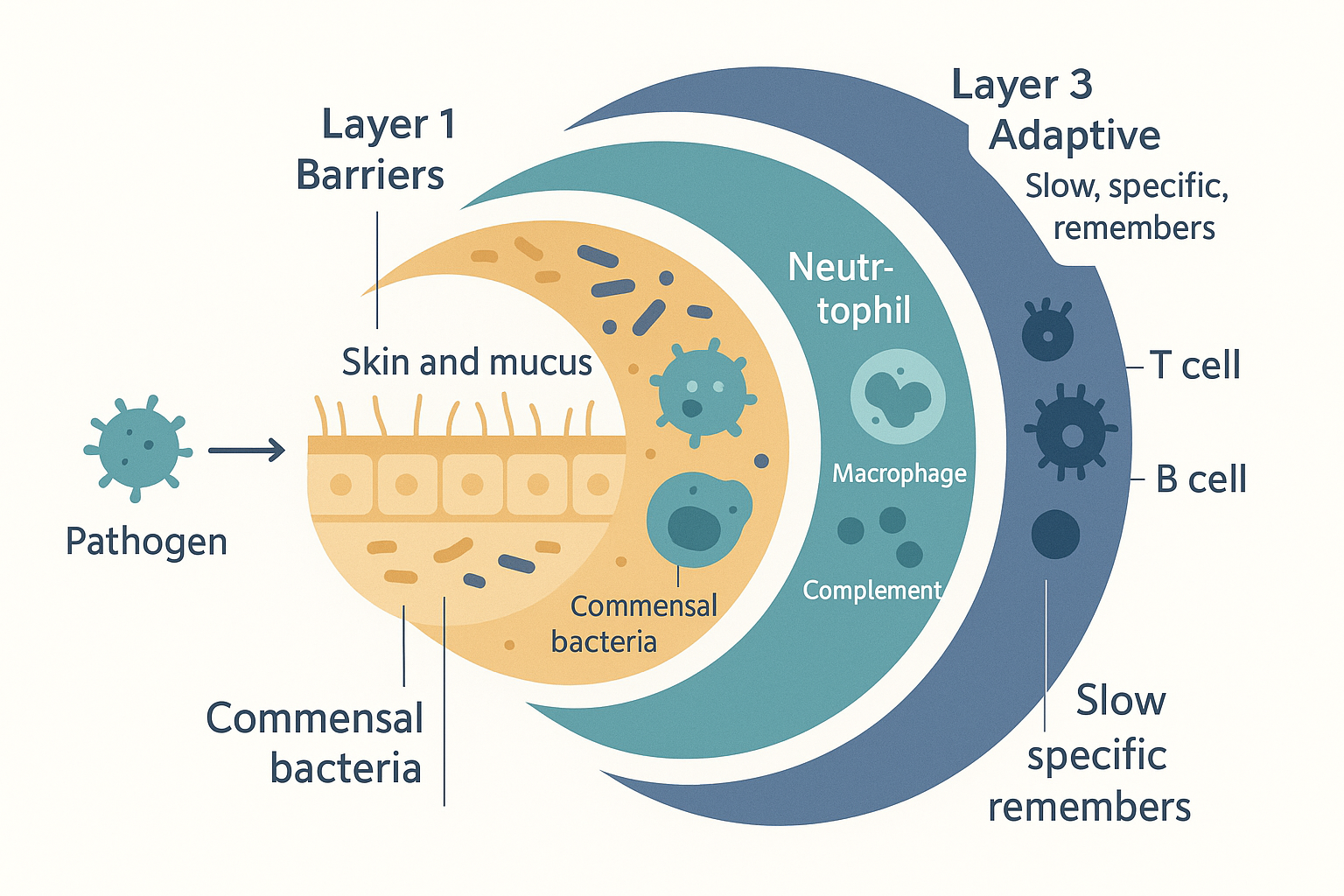 The three-layer model: barriers exclude most threats, fast innate immunity handles those that break through, and slow but specific adaptive immunity deals with what the innate system cannot — and remembers it for next time.
A note on the headline distinction, because it organises everything that follows:
| Innate immunity | Adaptive immunity | |
|---|---|---|
| Speed | Minutes–hours | Days (first exposure), hours (later) |
| Specificity | Broad categories (e.g. "bacterial wall") | One exact molecular target |
| How its sensors arise | Germline-encoded — fixed, inherited | Somatically generated — randomly assembled in each cell during your lifetime |
| Number of distinct targets | Hundreds of patterns | Billions of unique specificities |
| Memory | None | Yes — the defining feature |
| Main cells | Neutrophils, macrophages, dendritic cells, NK cells, mast cells | T lymphocytes, B lymphocytes |
| Main soluble weapons | Complement, interferons, cytokines | Antibodies |
Keep this table in mind. Most of the page is just filling in its rows.
Layer 1 — barriers: defence by exclusion
The cheapest defence is never letting the threat in. Long before any immune cell is involved, the body presents a set of physical and chemical barriers that stop the overwhelming majority of microbes.
- Skin — a dense, dry, constantly shedding sheet of dead keratin-filled cells. Most microbes simply cannot penetrate intact skin; this is why a breach (a cut, a burn, a needle) is the classic route of infection. It is also mildly acidic (the "acid mantle", pH ~5), which discourages bacterial growth.
- Mucous membranes — the wet linings of the airways, gut, and urogenital tract, where the body's surface is necessarily thin and permeable (it has to absorb air and food). These are defended by mucus, a sticky gel that traps incoming particles, and in the airways by cilia — microscopic beating hairs that sweep the trapped debris up and out (the "mucociliary escalator"). Coughing, sneezing, tears, and the constant flushing of urine are all mechanical clearance.
- Chemical defences — secretions laced with antimicrobial molecules. Lysozyme (an enzyme in tears, saliva, and mucus that dissolves bacterial cell walls), stomach acid (pH 1.5–3.5, a chemical furnace that kills most swallowed microbes — see digestion), defensins (small antimicrobial peptides), and bile.
- The commensal microbiota — the bacteria already living on and in you, especially in the gut. By occupying the available surface and consuming the available nutrients, they deny incoming pathogens a foothold — a phenomenon called colonisation resistance. This is a living barrier, and it is why broad-spectrum antibiotics, by clearing it, invite opportunistic infections like C. difficile.
Barriers are not glamorous, but they do most of the work. The rest of the immune system is what happens when a barrier is breached.
Layer 2 — innate immunity: the fast response
Once something foreign is inside the tissue, the innate immune system engages within minutes. Its job is to recognise that something is wrong using a small, fixed set of sensors, then to kill, contain, and raise the alarm. It cannot identify the specific invader — it works in broad categories — but it is fast and it is always ready.
How the innate system recognises threat: patterns, not specifics
The innate system does not recognise individual pathogens. Instead it recognises molecular patterns that are characteristic of whole classes of threat — and, crucially, that are absent from healthy human cells. There are two kinds of danger signal, both introduced on the inflammation page:
- PAMPs — pathogen-associated molecular patterns. Molecular signatures of invaders: the LPS (lipopolysaccharide) of gram-negative bacterial walls, the flagellin of bacterial tails, double-stranded RNA (a hallmark of viral replication), fungal cell-wall sugars. These are molecules microbes cannot easily abandon because they are structurally essential — which is why they make reliable targets.
- DAMPs — damage-associated molecular patterns. Molecules released by your own cells when they die violently or are injured — ATP, DNA, or uric acid spilling out where they should never be. DAMPs let the system respond to damage (a crush injury, a burn, dying tumour cells) even with no infection present.
These patterns are detected by pattern-recognition receptors (PRRs) — hard-wired sensors on and inside innate immune cells. The most famous family is the Toll-like receptors (TLRs), a set of about ten sensors each tuned to a different pattern: TLR4 detects bacterial LPS, TLR3 detects viral double-stranded RNA, TLR5 detects flagellin, and so on. When a PRR catches its pattern, it triggers the internal relay — converging on the master switch NF-κB — that launches inflammation (the full mechanism is on the inflammation page: NF-κB → cytokines TNF-α, IL-1β, IL-6 → vasodilation, leaky vessels, recruitment of more cells). Inflammation is the innate system's coordinated tissue response. This page is the organisation behind it.
The complement system: a soluble, self-assembling weapon
Before describing the cells, one purely chemical innate weapon deserves its own section because it is so often glossed over: complement. It is a cascade of about 30 blood proteins, made by the liver and circulating permanently in an inactive form, that "complements" the cells' work. When triggered, they activate one another in a chain reaction — each cleaving and activating the next — that amplifies a tiny initial signal into a large response within seconds.
Complement can be triggered three ways (the classical pathway, set off by antibodies bound to a target — a bridge from the adaptive system; the lectin pathway, set off by mannose sugars on microbial surfaces; and the alternative pathway, which ticks over spontaneously and is held in check only on self surfaces). All three converge on cleaving the central protein C3, and from there complement does three useful things:
- Opsonisation — it coats the microbe in fragments (chiefly C3b) that act as "handles", making it far easier for phagocytes to grab and eat. (Opsonise, from the Greek for "to prepare as food", is exactly the right image: complement seasons the bacterium so the eater can grip it.)
- Inflammation — released fragments (C3a, C5a) are potent chemoattractants, forming a chemical trail that recruits and guides neutrophils to the site.
- Direct lysis — the terminal components assemble into the membrane attack complex (MAC), a ring that punches a physical pore through the microbe's membrane, letting its contents leak out so it bursts.
flowchart TD
    T1["Classical<br/>(antibody-bound target)"] --> C3
    T2["Lectin<br/>(microbe sugars)"] --> C3
    T3["Alternative<br/>(spontaneous, on microbe surface)"] --> C3
    C3["Cleave C3 → C3a + C3b<br/>(central amplification step)"] --> OPS["C3b coats microbe<br/>= OPSONISATION (handles for phagocytes)"]
    C3 --> INF["C3a / C5a<br/>= recruit + guide neutrophils"]
    C3 --> MAC["Terminal components assemble<br/>MEMBRANE ATTACK COMPLEX → pore → lysis"]
The innate cells: the cast
The innate response is carried out by a set of white blood cells (leukocytes), most of them descended from a common myeloid precursor in the bone marrow. Know them by what they do:
- Neutrophils — the first responders and the most numerous white blood cell (50–70% of those in your blood). They swarm a site within hours, engulf and destroy bacteria, and deploy a startling repertoire: phagocytosis, a "respiratory burst" of bactericidal reactive oxygen, and NETs (neutrophil extracellular traps — webs of their own DNA flung out to ensnare microbes). They are short-lived kamikaze cells; pus is largely spent neutrophils. They are the dominant cell of acute inflammation.
- Monocytes / macrophages — the "big eaters" (literally what macrophage means). Monocytes circulate in the blood; when they enter tissue they mature into macrophages. They are professional phagocytes that devour pathogens and cellular debris, but they are far more than garbage disposal: they are sentinels stationed in every tissue, the major source of the alarm cytokines, and the cells that later switch from "attack" to "repair" mode to orchestrate healing. Some are long-lived tissue residents with their own names (microglia in the brain, Kupffer cells in the liver).
- Dendritic cells — the bridge to the adaptive system, important enough that they get their own section below. Their day job is patrolling the barriers, sampling everything, and carrying what they find back to the lymph nodes.
- Natural killer (NK) cells — the anti-viral and anti-tumour innate cell. Confusingly named (they are lymphocytes by lineage but innate by behaviour), they kill infected and cancerous host cells rather than free microbes. Their logic is elegantly inverted: most healthy cells display a "self" molecule called MHC class I (explained below) on their surface as a passport. Viruses and tumours often down-regulate MHC class I to hide from T cells — and NK cells are built to kill any cell showing "missing self". So the very trick a virus uses to evade one arm of immunity exposes it to the other.
- Mast cells, basophils, eosinophils — the inflammatory and anti-parasite granulocytes. Mast cells sit in tissues packed with granules of histamine and release them explosively to drive vasodilation and leakiness (the same histamine antihistamines block; the central player in allergy, below). Basophils are their circulating cousins. Eosinophils specialise in large parasites (worms) too big to engulf, attacking them with toxic granule proteins; they are also heavily involved in allergic disease.
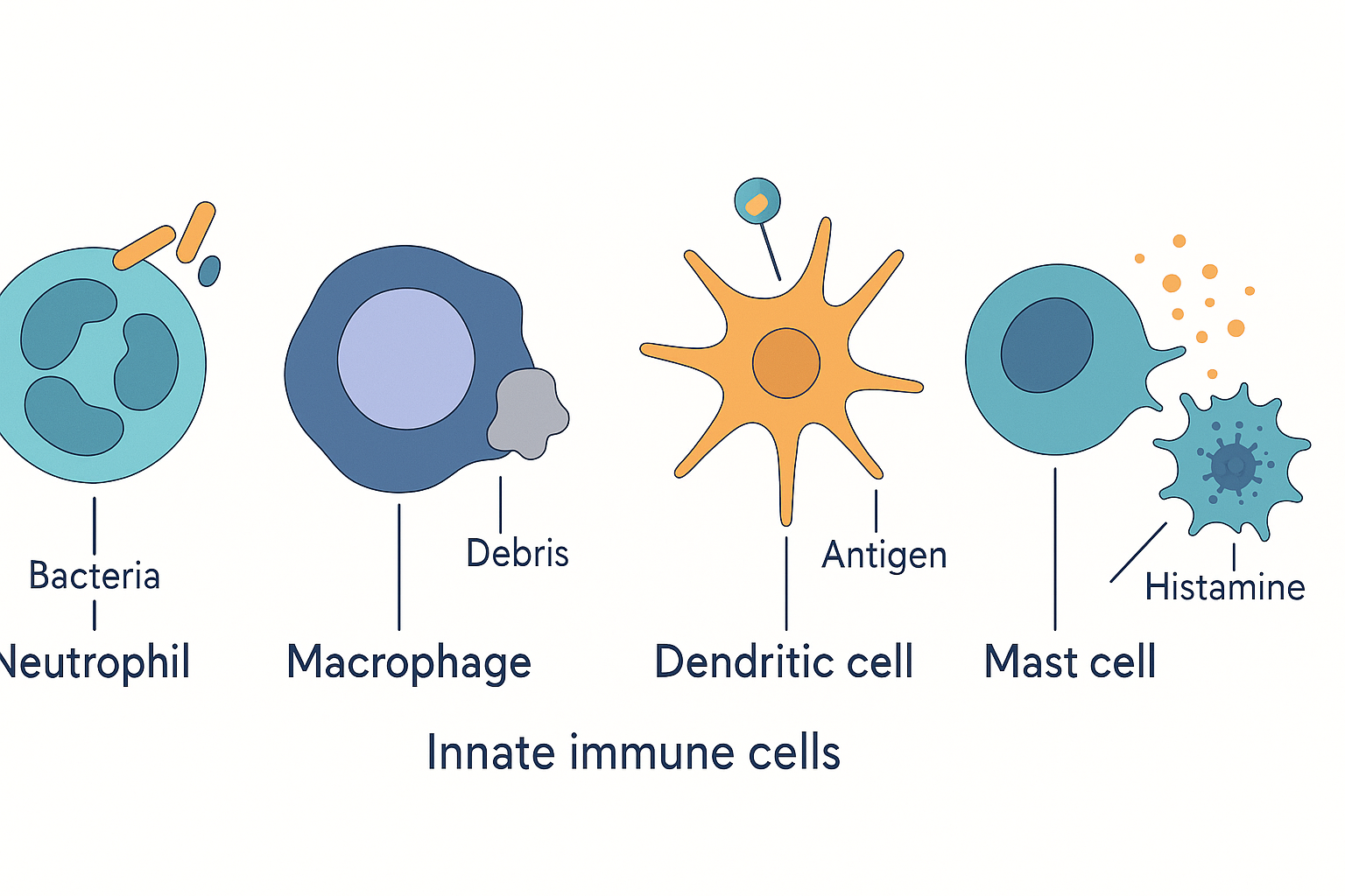 The innate cast: neutrophils (first-responder bacteria-killers), macrophages (eaters and sentinels), dendritic cells (the bridge to adaptive immunity), NK cells (kill virus-infected and tumour cells via "missing self"), and mast cells (explosive histamine release driving inflammation and allergy).
Interferons: the anti-viral broadcast
One more innate weapon, specific to viruses. When a cell detects it is infected (its internal PRRs sense viral RNA), it secretes interferons (IFN) — signalling proteins that "interfere" with viral replication. Type I interferons (IFN-α and IFN-β) act as a warning broadcast to neighbouring cells, telling them to switch on hundreds of anti-viral genes and enter a defensive state that makes them hostile to viral copying before they are even infected. Interferons also sharpen NK-cell killing. They are why a viral infection produces such systemic, achy, feverish malaise — and interferon-γ (IFN-γ), made later by adaptive cells, is the key signal that ramps up macrophage killing (a Th1 cytokine, below).
The bridge: dendritic cells and antigen presentation
Here is the single most important hand-off in immunology — the moment the fast innate system recruits the slow adaptive one. It is worth slowing down for.
The dendritic cell (DC) is an innate cell whose whole purpose is to be a messenger. It sits in the barrier tissues — skin, gut lining, airways — with long branching arms (dendrites) constantly sampling its surroundings by engulfing bits of everything around it. When it engulfs a pathogen in a context of danger (i.e. its PRRs are also firing because PAMPs are present), the dendritic cell matures: it stops sampling, packages fragments of the pathogen, and migrates through the lymphatic vessels to the nearest lymph node — a meeting hall where adaptive lymphocytes congregate.
To understand what it does there, you need one central concept: antigen presentation via MHC.
- An antigen is any molecular fragment that the adaptive immune system can specifically recognise — typically a short piece of protein (a peptide) from a pathogen.
- MHC (major histocompatibility complex) molecules are the body's display platforms: surface proteins whose job is to hold up a peptide fragment for T cells to inspect. Think of MHC as a hand holding a sample up to be checked. (In humans these are also called HLA, human leukocyte antigens — the molecules matched in organ transplants.) There are two classes, and the distinction is fundamental:
- MHC class I is on almost every cell in your body. It continuously displays peptides from inside the cell — a real-time readout of what proteins the cell is making. A healthy cell shows only "self" peptides; a virus-infected or cancerous cell inevitably shows fragments of viral or mutant protein. MHC class I is inspected by cytotoxic (CD8+) T cells, which kill any cell displaying foreign peptide. It is the body's universal "show me your insides" passport system.
- MHC class II is only on professional antigen-presenting cells (APCs) — dendritic cells, macrophages, and B cells. It displays peptides the cell engulfed from outside itself, and it is inspected by helper (CD4+) T cells. It is the "look what I found out there" channel.
So the dendritic cell, arriving in the lymph node bearing pathogen fragments on its MHC class II, is presenting evidence to the helper T cells: here is what I found at the barrier, and here is the danger context that came with it. That presentation is the spark that ignites the entire adaptive response. No presentation, no adaptive immunity — which is exactly why dendritic cells are described as the bridge.
Layer 3 — adaptive immunity: the slow specialists
The adaptive immune system is built from a single cell type — the lymphocyte — in two great families: T cells and B cells. (Both are made in the bone marrow; T cells are then schooled in the thymus, B cells mature in the marrow — hence the names, T for thymus, B originally for the bursa in birds where they were discovered.) Adaptive immunity has two defining properties that the innate system lacks: specificity (each lymphocyte recognises one exact molecular target) and memory. Both fall out of one beautiful mechanism.
Clonal selection: how one-in-a-billion specificity works
Burnet's 1957 insight. The body does not design a receptor to fit each new pathogen — that would be impossible, since pathogens are unpredictable. Instead it builds, in advance, an enormous repertoire of lymphocytes, each carrying a single, randomly generated receptor of a different specificity — on the order of a billion distinct specificities, ready before any infection. The overwhelming majority of these will never meet their match in a lifetime. When a pathogen does arrive, it acts as a selector: of the billion lymphocytes, the rare few whose receptor happens to fit a fragment of that pathogen are activated, and they proliferate — divide rapidly into a large army of identical clones, all specific for the invader. This is clonal selection: the antigen does not instruct; it selects and amplifies a pre-existing clone.
This single mechanism explains three things at once: - Specificity — only the matching clone expands. - Memory — some of the expanded clone persist long-term as memory cells, so the clone is now large and pre-armed for next time. - Self-tolerance — clones that would attack your own tissues are deleted early (see tolerance, below), so the surviving repertoire is pointed outward.
V(D)J recombination: where the billion specificities come from
How can the genome encode a billion different receptors when it has only ~20,000 genes? It does not store them — it assembles them randomly. The receptor's recognition region is not written as a single gene but split into gene segments filed in three groups, named V (variable), D (diversity), and J (joining). As each lymphocyte matures, a dedicated cut-and-paste enzyme system (the RAG recombinase) selects one segment from each group at random and stitches them together to build that cell's unique receptor gene — and adds further randomness by inserting or trimming a few extra nucleotides at each join. This process is called V(D)J recombination.
The combinatorial maths is the point: a few dozen V, a handful of D, a few J segments, multiplied together and randomised at the joins, generate billions of distinct receptors from a modest stretch of DNA. Each lymphocyte runs the lottery once, gets one receptor for life, and passes that specificity to all its clones. (B cells later refine this: after activation they undergo somatic hypermutation, deliberately mutating the receptor gene further and selecting the highest-affinity variants — affinity maturation — which is why antibodies get better over the course of a response.) This is what "somatically generated specificity" in the comparison table means: the diversity is created in your body's cells during your lifetime, not inherited.
B cells and antibodies: humoral immunity
A B cell's receptor is a membrane-bound antibody. When a B cell's antibody binds its matching antigen and the B cell receives a confirmatory "go" signal from a helper T cell (the two-signal safety check, below), the B cell activates, proliferates, and differentiates into a plasma cell — an antibody factory that secretes thousands of copies of its antibody per second into the blood and lymph. This is humoral immunity (from "humour", the old word for body fluids): defence carried by soluble molecules, Ehrlich's chemical arm.
An antibody (also called an immunoglobulin, Ig) is a Y-shaped protein. The two tips of the Y are the variable, antigen-binding ends — the part built by V(D)J recombination, which clamps onto the specific target. The stem of the Y (the constant region) is the "handle" that the rest of the immune system grabs, and it comes in different versions that determine what the antibody does. Antibodies do not usually kill anything directly; they mark, neutralise, and recruit:
- Neutralisation — by physically coating a virus or toxin, they block it from binding host cells.
- Opsonisation — they coat a microbe so phagocytes (which have receptors for the antibody stem) can grip and eat it.
- Complement activation — bound antibodies trigger the classical complement pathway.
- Recruitment — the stem flags the target for NK cells and other killers.
There are five antibody classes, distinguished by their stem (constant) region, each with a division of labour:
| Class | Where / form | What it does |
|---|---|---|
| IgG | Most abundant in blood (~75% of serum antibody); a single Y | The workhorse of the secondary response: high-affinity, long-lived, opsonises, neutralises, activates complement. The only class that crosses the placenta, giving newborns passive maternal immunity. |
| IgA | Secretions — saliva, tears, breast milk, gut and airway mucus; usually a dimer (two Ys joined) | Mucosal defence — the dominant antibody at the barriers, neutralising pathogens before they cross. Vast amounts are made in the gut; breast-milk IgA protects the infant gut. |
| IgM | A large pentamer (five Ys joined); stays in the bloodstream | The first responder of the antibody world — secreted early in a primary response before affinity maturation. Its ten binding sites make it a powerful complement activator despite individually weak binding. |
| IgE | Trace amounts; bound to mast cells and basophils | Anti-parasite — and the villain of allergy. When its target cross-links IgE on a mast cell, the cell explodes with histamine. The IgE/mast-cell axis is the engine of hay fever, asthma, and anaphylaxis (below). |
| IgD | On the surface of naïve B cells; little secreted | Largely a B-cell receptor during maturation; its circulating role remains the least understood. |
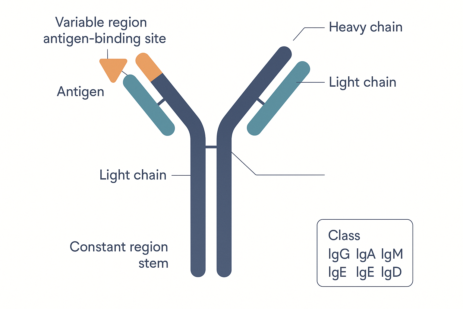 Antibody architecture: the two variable tips (built by V(D)J recombination) clamp the specific antigen; the constant stem is the handle that recruits phagocytes, complement and killer cells — and whose version defines the five classes IgG, IgA, IgM, IgE, IgD.
T cells: cell-mediated immunity
Where B cells fight extracellular threats with secreted antibodies, T cells handle the threats antibodies cannot reach — chiefly cells that are already infected from within (viruses hide inside host cells) — and they direct the rest of the immune system. A T cell's receptor (the TCR) does not recognise free antigen; it only recognises a peptide presented on an MHC molecule (the reason antigen presentation is so central). T cells come in subsets defined by a surface marker and a job:
- Cytotoxic T cells (CD8+) — the assassins of cell-mediated immunity. The CD8 co-receptor binds MHC class I, so these cells inspect the "show me your insides" display on every body cell. When a CD8+ T cell finds a cell presenting foreign peptide (a virus-infected or cancerous cell), it kills it precisely — punching it with perforin (a pore-former) and injecting granzymes (enzymes that trigger the target's own suicide programme, apoptosis). This is how the body eliminates infected cells with the contents intact, avoiding the mess of bursting them open.
- Helper T cells (CD4+) — the conductors. The CD4 co-receptor binds MHC class II, so these cells inspect what the antigen-presenting cells bring back. Helper T cells kill nothing directly; instead they secrete cytokines that orchestrate everyone else — licensing B cells to make antibodies, activating macrophages to kill harder, and recruiting other cells. They are the central coordinators, which is precisely why HIV, by destroying CD4+ T cells, is so devastating: knock out the conductors and the whole orchestra falls apart.
- Regulatory T cells (Treg) — the brakes. A specialised CD4+ subset whose entire job is to suppress immune responses — to switch the system off when the threat is cleared and, critically, to enforce tolerance toward self and harmless antigens (food, commensal bacteria). They are introduced here and feature heavily in tolerance and autoimmunity below.
Helper T-cell subsets: the Th1/Th2/Th17/Treg decision
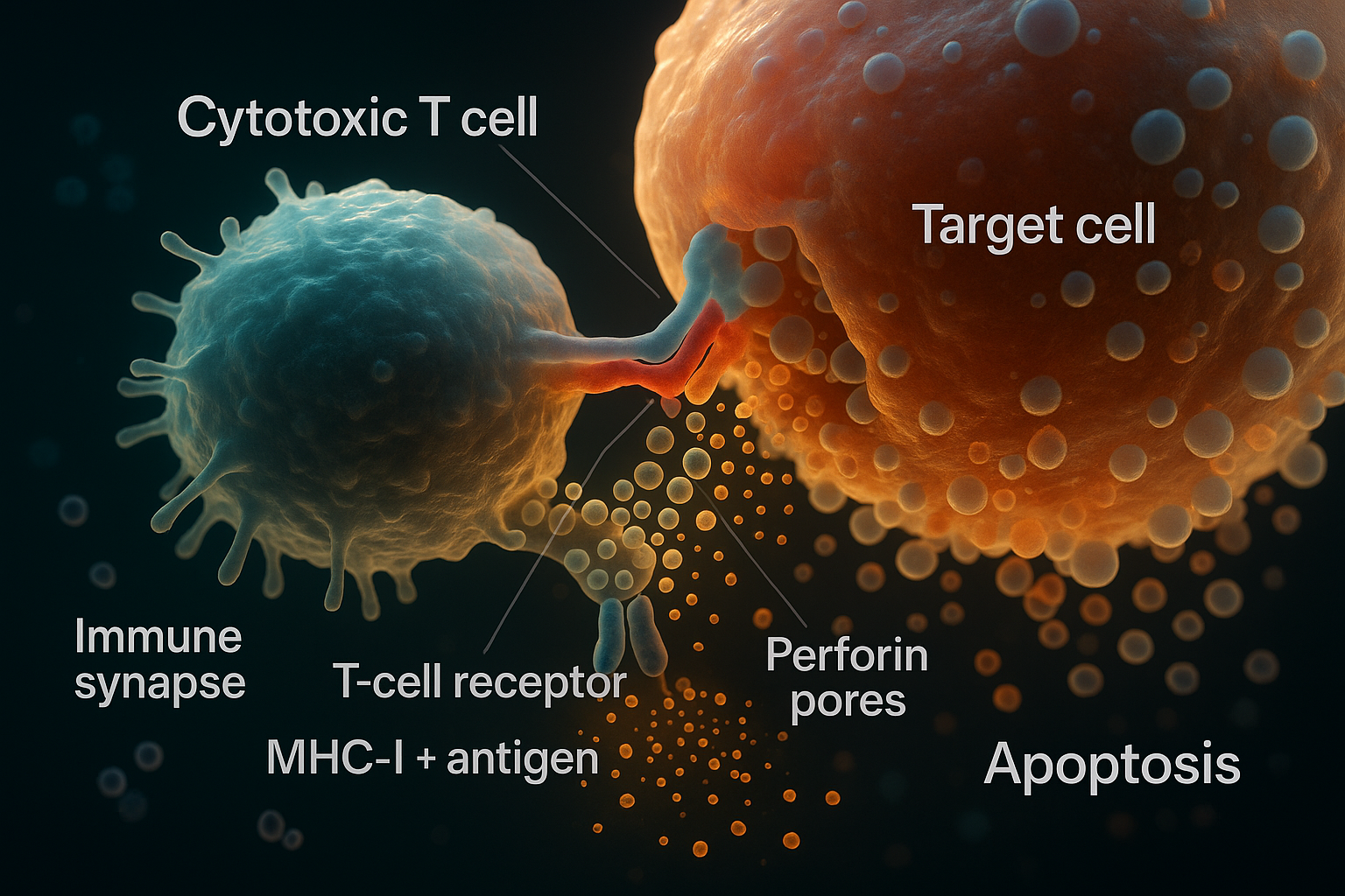 The assassin at work: a cytotoxic (CD8+) T cell forms an immune synapse with a target cell whose MHC-I is displaying foreign peptide, then fires perforin (which punches pores in the target membrane) and granzymes (which trigger the target’s own apoptosis) — destroying the infected or cancerous cell cleanly from the inside.
A naïve helper (CD4+) T cell — one that has never yet met its antigen — when first activated, must choose what kind of response to coordinate — and it chooses based on the cytokines in the environment, which the innate system set according to the type of threat it detected. This is one of the most important regulatory branch-points in immunology, because it tailors the response to the pathogen — and because getting the choice wrong underlies allergy and autoimmunity.
- Th1 — coordinates defence against intracellular threats (viruses, bacteria living inside cells). Driven by IL-12 and IFN-γ; its signature output is IFN-γ, which supercharges macrophage killing and CD8+ T cells. The "cell-mediated" programme.
- Th2 — coordinates defence against large parasites (worms) and drives the antibody/allergy axis. Its cytokines (IL-4, IL-5, IL-13) recruit eosinophils, activate mast cells, and push B cells to make IgE. An inappropriate Th2 response to a harmless antigen is allergy.
- Th17 — coordinates defence against extracellular bacteria and fungi at barriers; its cytokine IL-17 recruits neutrophils to mucosal surfaces. Important in gut immunity — and over-active Th17 responses feature in autoimmune diseases like psoriasis and inflammatory bowel disease.
- Treg — the suppressive choice (above): instead of mounting a response, enforce tolerance.
The Th1/Th2 balance is a recurring theme in immunology and in biohacking discussion: the two arms are partly mutually inhibitory (IFN-γ suppresses Th2; IL-4 suppresses Th1), so the system tends to polarise, and a chronic skew one way or the other shapes which diseases a person is prone to.
flowchart TD
    APC["Dendritic cell presents antigen<br/>+ sets cytokine context by threat type"] --> NAIVE["Naive CD4+ helper T cell<br/>(must choose a programme)"]
    NAIVE -->|"IL-12, IFN-γ<br/>(intracellular: virus)"| TH1["Th1 → IFN-γ<br/>activate macrophages + CD8 killers"]
    NAIVE -->|"IL-4<br/>(parasites / allergens)"| TH2["Th2 → IL-4,5,13<br/>eosinophils, mast cells, IgE"]
    NAIVE -->|"IL-6, TGF-β<br/>(extracellular bacteria/fungi)"| TH17["Th17 → IL-17<br/>recruit neutrophils to barriers"]
    NAIVE -->|"TGF-β, retinoic acid<br/>(harmless / self)"| TREG["Treg → IL-10, TGF-β<br/>SUPPRESS, enforce tolerance"]
    TH2 -.->|"misfires on harmless antigen"| ALLERGY["ALLERGY"]
    TH17 -.->|"misfires on self"| AUTO["AUTOIMMUNITY"]
Putting the activation sequence together
We can now trace the whole adaptive response as a single chain of events, from breach to antibody:
sequenceDiagram
participant P as Pathogen (at barrier)
participant DC as Dendritic cell
participant Th as Helper CD4+ T cell
participant B as B cell
participant Tc as Cytotoxic CD8+ T cell
P->>DC: engulfed in danger context (PAMPs)
DC->>DC: mature, migrate to lymph node
DC->>Th: present peptide on MHC-II (+ co-stimulation)
Note over Th: clonal selection — matching clone activates + proliferates,<br/>chooses Th1/Th2/Th17 programme
Th->>B: "go" signal (signal 2) + cytokines
Note over B: B whose antibody fits antigen activates →<br/>plasma cells secrete antibody; some become memory B
Th->>Tc: help / cytokines
Note over Tc: CD8 clone matching MHC-I-presented peptide expands →<br/>kills infected cells; some become memory T
B-->>P: antibodies neutralise / opsonise
Tc-->>P: kill infected host cells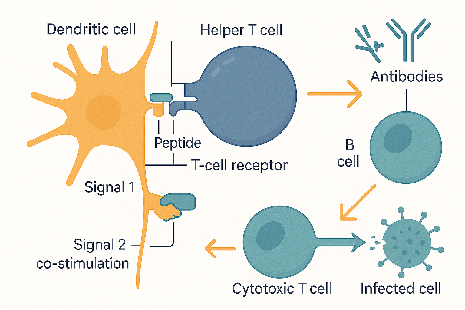 The hand-off that ignites adaptive immunity: a dendritic cell presents antigen on MHC to a helper T cell's receptor (signal 1) plus a co-stimulation handshake (signal 2); the activated helper T cell then licenses B cells to make antibodies and helps cytotoxic T cells kill infected cells.
A crucial safety detail visible in that diagram: T and B cells require two signals to activate. Signal 1 is the specific match (TCR to peptide-MHC). Signal 2 is a separate co-stimulation handshake that the antigen-presenting cell only provides when it has also sensed genuine danger (PAMPs). A lymphocyte that receives signal 1 without signal 2 — i.e. it matches an antigen, but no danger was present — is not activated; it is switched off (made anergic) or deleted. This is a core mechanism of peripheral tolerance (below): it prevents the system attacking harmless self-antigens just because they happen to match a receptor.
Where immune cells are made and trained: the lymphoid organs
All of this machinery is built, schooled, and deployed in a dedicated set of organs, split into two tiers. (This whole section sets up the sibling pages on the thymus and the lymphatic system.)
Primary lymphoid organs — where lymphocytes are made and educated:
- Bone marrow — the birthplace of all blood cells. A population of haematopoietic stem cells here gives rise to every red cell, platelet, and white cell, via two great lineages: the myeloid line (neutrophils, monocytes/macrophages, dendritic cells, mast cells, eosinophils) and the lymphoid line (T cells, B cells, NK cells). It is also where B cells mature and are tested for self-reactivity.
- Thymus — a small gland behind the breastbone, dedicated entirely to T-cell education (the T in T cell). Immature T cells migrate here from the marrow and run a brutal two-part exam — positive selection (can your receptor recognise self-MHC at all? if not, useless — die) and negative selection (does your receptor bind self too strongly? if so, dangerous — die). Only ~2% pass both and are released as mature, self-tolerant T cells. The thymus is central to two later sections: central tolerance, and immunosenescence — because the thymus shrinks with age (involution), so T-cell training winds down across life. This is the heart of the forthcoming thymus page and the rationale behind thymic peptides like thymalin and vilon.
Secondary lymphoid organs — where lymphocytes meet antigen and respond:
- Lymph nodes — small bean-shaped filters strung along the lymphatic vessels (the lymphatic system), positioned to catch antigen draining from the tissues. They are the meeting halls: dendritic cells arrive here bearing antigen and present it to the dense population of T and B cells waiting inside. This is where clonal selection physically happens, and why nodes swell during an infection — they are packed with proliferating clones. (The lymphatics are also how those dendritic cells travelled in the first place — see the lymphatic system.)
- Spleen — the same function as a lymph node, but filtering the blood rather than the lymph: it catches blood-borne pathogens and also clears old red blood cells.
- MALT / GALT — mucosa- and gut-associated lymphoid tissue: the immune outposts embedded directly in the barrier linings, where most pathogens actually arrive. The gut's GALT (including the Peyer's patches) is the largest single concentration of immune tissue in the body — fitting, since the gut is the surface most exposed to the outside world.
flowchart TD
    HSC["Bone marrow<br/>haematopoietic stem cell"] --> MYE["Myeloid line:<br/>neutrophils, monocytes/macrophages,<br/>dendritic cells, mast cells, eosinophils"]
    HSC --> LYM["Lymphoid line:<br/>T cells, B cells, NK cells"]
    LYM --> BCELL["B cells mature in marrow<br/>(tested for self-reactivity)"]
    LYM --> TCELL["T cells migrate to THYMUS<br/>positive + negative selection<br/>(~2% survive, self-tolerant)"]
    BCELL --> SEC
    TCELL --> SEC["Secondary organs<br/>(meet antigen)"]
    SEC --> LN["Lymph nodes<br/>(filter lymph)"]
    SEC --> SP["Spleen<br/>(filter blood)"]
    SEC --> GALT["GALT / MALT<br/>(barrier outposts, largest mass)"]
Immunological memory and vaccination
We can now explain the single most consequential property of adaptive immunity — and the basis of all of Jenner's legacy.
When a clone expands to fight an infection, most of the expanded cells are short-lived effector cells that die once the threat is cleared. But a fraction persist for years or decades as memory cells — memory B cells and memory T cells. The clone that was one-in-a-billion before the infection is now large, pre-selected, and primed. This converts a slow first response into a fast second one:
- The primary response (first exposure) is slow — days to find and expand the rare matching clone — and the antibody it produces is initially the lower-affinity IgM, switching later to IgG.
- The secondary response (re-exposure to the same antigen) is faster, larger, and higher-quality: the memory clone is already abundant, already affinity-matured, and immediately produces large amounts of high-affinity IgG. The pathogen is often neutralised before it can cause symptoms. This is what we mean by being "immune."
flowchart LR
    E1["1st exposure"] --> P["Primary response:<br/>slow (days), small, IgM→IgG"]
    P --> MEM["Memory B + T cells persist<br/>(clone now large + primed)"]
    MEM --> E2["2nd exposure (same antigen)"]
    E2 --> S["Secondary response:<br/>fast (hours), large, high-affinity IgG<br/>→ often no symptoms"]
Vaccination is this mechanism, deliberately triggered with a safe version of the threat — a killed or weakened microbe, a fragment of it, or (in mRNA vaccines) the genetic instructions for the body to briefly make one harmless piece of it. The immune system mounts a primary response and lays down memory without the danger of the real disease, so that the first encounter with the live pathogen is met by an already-primed secondary response. Jenner did exactly this with cowpox in 1796 without understanding any of it; we now do it on purpose, and increasingly with programmable precision.
Self versus non-self: tolerance and what goes wrong
Everything so far assumes the immune system attacks foreign things and leaves you alone. But a system that randomly generates a billion receptors will inevitably generate many that fit your own tissues. Preventing those from causing harm — self-tolerance — is as important as recognising the foreign, and its failures define a whole class of disease.
Tolerance is enforced in two tiers:
- Central tolerance — the culling described above, during development. In the thymus, T cells whose receptors bind self-peptides too strongly are deleted (negative selection); in the bone marrow, self-reactive B cells are likewise removed. This eliminates the most dangerous self-reactive clones before they ever enter circulation.
- Peripheral tolerance — the backup, out in the body, catching self-reactive cells that slipped through. Its mechanisms include the two-signal rule (a lymphocyte that matches an antigen but receives no "danger" co-stimulation is switched off rather than activated — see above) and, above all, the regulatory T cells (Tregs) that actively suppress responses against self and harmless antigens.
When tolerance fails or the system is mis-calibrated, you get the three great failure modes:
- Autoimmunity — the immune system attacks self. A self-reactive clone escapes tolerance and treats a body tissue as foreign: the pancreatic beta cells in type 1 diabetes, the thyroid in Hashimoto's, the joints in rheumatoid arthritis, myelin in multiple sclerosis. This is the system being too aggressive against the wrong target — and it underlies a large share of the chronic inflammation the previous page described as having an "autoimmune process" among its persistent triggers.
- Allergy / hypersensitivity — the immune system attacks harmless things. The classic (Type I) form is the IgE/mast-cell axis: an inappropriate Th2 response treats a harmless antigen (pollen, peanut, cat dander) as a parasite, making IgE against it. The IgE arms the mast cells; on re-exposure, the allergen cross-links that IgE and the mast cells detonate, dumping histamine — producing the runny nose, hives, or, when body-wide, the catastrophic vasodilation of anaphylaxis. Allergy is the immune system correctly executing a Th2/IgE programme against an incorrectly chosen target. (The hygiene hypothesis proposes that a modern childhood with too few microbial challenges under-trains the regulatory arm, skewing toward Th2 and allergy — one of the strongest arguments for the microbiome's role in immune calibration.)
- Immunodeficiency — the immune system is too weak. Either inherited (e.g. defects in particular cells) or acquired (HIV destroying CD4+ helper T cells; chemotherapy or immunosuppressant drugs; severe malnutrition). The result is vulnerability to infections a normal system would shrug off — and a reminder that the goal is never simply "less immune activity."
These three together make the central point of the whole page concrete: immune health is calibration, not amplitude. Too strong against the wrong target is autoimmunity; too strong against the harmless is allergy; too weak is immunodeficiency. The well-regulated middle is the target.
The regulation layer and systems links
Finally, the connective tissue — how the immune system talks to itself and to the rest of the body, and where the biohacking levers actually sit.
Cytokines: the messaging layer
The immune system has no wiring of its own; it coordinates chemically, through cytokines — small signalling proteins that act as the immune system's hormones, letting one cell instruct another. They are the language in which the whole system above is actually spoken. A working vocabulary:
- TNF-α, IL-1β, IL-6 — the pro-inflammatory trio (full treatment on the inflammation page): made by activated innate cells, they drive local inflammation, fever, and the liver's acute-phase response.
- Interferons (IFN-α/β anti-viral; IFN-γ the Th1 macrophage-activator) — covered above.
- The interleukins (IL-…) — a large numbered family that does most of the directing: IL-12 pushes Th1, IL-4 pushes Th2, IL-17 is the Th17 output, and so on.
- IL-10 and TGF-β — the anti-inflammatory / suppressive cytokines, the Tregs' main tools for switching responses off and enforcing tolerance. They are the chemical "stand down" order, the counterpart to the resolution programme on the inflammation page.
The balance between the pro-inflammatory and the suppressive cytokines is, in effect, the immune system's volume control — and it is what most "immunomodulatory" interventions are nudging.
Neuro-immune links: the brain has a dial on immunity
The immune system is not autonomous; it is wired to the nervous system. The key link, developed in full on the autonomic nervous system page, is the cholinergic anti-inflammatory reflex: sensory fibres of the vagus nerve detect inflammatory cytokines in the body and report to the brainstem; the brain responds by sending efferent vagal signals that, via the spleen, cause acetylcholine release that directly suppresses TNF-α production by macrophages. This is a literal neural brake on inflammation — the mechanism behind why high vagal tone, slow breathing, and meditation are measurably anti-inflammatory, and why chronic stress (sympathetic dominance, dysregulated cortisol) is pro-inflammatory. The brain and immune system are two-way wired.
The gut as an immune organ
The largest mass of immune tissue in the body sits in the gut wall (GALT), in constant negotiation with the microbiome. This is where much of the immune system is trained: the commensal bacteria continuously educate the local immune cells — in particular driving the development of Tregs (the gut uses TGF-β and bacterial metabolites like the short-chain fatty acid butyrate to induce them) — teaching the system to tolerate food and friendly bacteria while staying ready for pathogens. A disrupted microbiome therefore means a mis-trained immune system, the mechanistic core of the link between gut health and both allergy and autoimmunity. And when the gut barrier leaks, the LPS that crosses drives exactly the TLR4 → NF-κB inflammation described at the top of this page — the through-line connecting the gut, the immune, and the inflammation pages into one story.
Nutrient cofactors: the raw materials
An immune system is built and run from molecular raw materials; deficiency degrades it. The ones with the strongest mechanistic basis:
- Vitamin D — acts almost as an immune hormone: its receptor is on most immune cells, and it both supports antimicrobial defence (it induces the antimicrobial peptide cathelicidin) and promotes the regulatory/tolerogenic side (favouring Tregs). Deficiency tracks with both more infection and more autoimmunity — fitting its role as a calibrator rather than a booster.
- Zinc — required for the function and development of essentially all immune cells, especially T cells; deficiency causes thymic atrophy and shrinks the lymphocyte pool. One of the most consistently immune-critical micronutrients.
- Vitamin A (as retinoic acid) — central to barrier (mucosal) integrity and, like vitamin D, to inducing gut Tregs and directing IgA responses; deficiency notoriously increases infection susceptibility.
- Vitamin C — concentrated in neutrophils, supports their oxidant killing and later clearance; a classic antioxidant cofactor for immune-cell function.
- Selenium and glutathione — the antioxidant defence that lets immune cells deploy reactive oxygen as a weapon without destroying themselves; glutathione status also influences the Th1/Th2 balance.
The honest framing: these correct deficiencies. A replete system is not improved by megadoses — the curve is a plateau, not a ramp — which is why "calibration, not amplitude" applies to nutrition too.
Immunosenescence: the ageing immune system
The immune system ages, and predictably — a process called immunosenescence. Two changes dominate. First, the thymus involutes: it begins shrinking from adolescence and is largely replaced by fat by old age, so the output of new, naïve T cells dwindles. The ageing immune system runs increasingly on its existing memory cells and struggles to mount fresh responses to novel threats — which is why the old are more vulnerable to new pathogens and respond less well to vaccines. Second, ageing brings a chronic, sterile, low-grade background inflammation — "inflammaging" (the inflammation page's chronic state) — a smouldering immune over-activity coexisting with under-effective specific defence: simultaneously over- and under-active, the calibration failing in both directions. This is the rationale behind interest in thymic and pineal restorative peptides — thymalin, vilon (see the Vilon deep dive), and epithalon — and the central theme the forthcoming thymus page will develop.
Putting it all together
- The immune system is a three-layer defence: barriers (skin, mucus, acid, commensals) exclude most threats; innate immunity (fast, germline-encoded, non-specific, no memory) handles breaches within hours; adaptive immunity (slow first time, somatically-generated specificity, long memory) deals with what the innate cannot — and remembers it.
- Innate immunity recognises broad patterns (PAMPs from microbes, DAMPs from damaged self) via fixed sensors (TLRs), launching inflammation (the inflammation page in full). Its weapons: phagocytes (neutrophils, macrophages), the complement cascade (opsonise, recruit, lyse), NK cells (kill "missing-self" infected/tumour cells), mast cells (histamine), and interferons (anti-viral).
- The dendritic cell bridges the two halves: it samples the barrier, and on detecting danger carries antigen to a lymph node and presents it on MHC to T cells — the spark of adaptive immunity. MHC class I ("show me your insides", on all cells, read by CD8+ killers); MHC class II ("look what I found", on antigen-presenting cells, read by CD4+ helpers).
- Adaptive immunity works by clonal selection on a pre-built repertoire of a billion specificities, generated by random V(D)J recombination. B cells become plasma cells secreting antibodies (humoral; five classes — IgG workhorse, IgA mucosal, IgM first-responder, IgE allergy/parasite, IgD). T cells are cytotoxic (CD8+) assassins of infected cells, helper (CD4+) conductors that choose a Th1/Th2/Th17/Treg programme by cytokine context, and Treg brakes.
- Lymphocytes are made and trained in primary organs (bone marrow = all blood cells + B-cell maturation; thymus = T-cell education) and meet antigen in secondary organs (lymph nodes, spleen, GALT/MALT).
- Memory cells convert a slow primary response into a fast, overwhelming secondary one — the basis of lasting immunity and of vaccination.
- Tolerance keeps the system pointed outward: central (thymic/marrow deletion of self-reactive clones) and peripheral (the two-signal rule, Tregs). Its failures are the three great modes: autoimmunity (too strong, wrong target), allergy (too strong, harmless target — the IgE/mast-cell axis), and immunodeficiency (too weak).
- It is all coordinated by cytokines (pro-inflammatory TNF-α/IL-1/IL-6 vs suppressive IL-10/TGF-β), wired to the brain via the vagal cholinergic anti-inflammatory reflex (ANS page), trained by the gut microbiome (GALT, butyrate-induced Tregs), supplied by nutrient cofactors (vitamin D, zinc, vitamin A, vitamin C, selenium, glutathione), and ages via thymic involution and inflammaging.
The single unifying idea: the immune system is not a force to be boosted but a network to be calibrated. Its genius is the layering — cheap exclusion, fast general defence, slow precise defence — and its discipline is self-restraint: a system that can generate a billion ways to attack must spend as much effort learning what not to attack. Health is the well-regulated middle, and almost every real lever (gut, vagal tone, sleep, deficiency correction) works by improving the regulation, not the volume.
Related Compounds & Deep Dives
Thymic & restorative peptides (immunosenescence)
- Thymalin — thymic peptide aimed at restoring T-cell output as the thymus involutes.
- Vilon & the Vilon deep dive — short peptide studied for immune/age-related regulatory effects.
- Vesugen — vascular/peptide bioregulator in the same Khavinson family.
- Pinealon & Epithalon deep dive — pineal peptides linked to the neuro-immune-ageing axis.
Barrier & mucosal immunity (IgA, gut training)
- Lactoferrin & Colostrum deep dive — passive immune factors and antimicrobial proteins that support the mucosal barrier.
- Lactoferrin, Colostrum — supply secretory IgA and iron-sequestering antimicrobial defence at the gut barrier.
- KPV Peptide — anti-inflammatory tripeptide acting on mucosal/innate inflammation.
- Probiotics, Akkermansia muciniphila, Butyrate — train the GALT and induce regulatory T cells via the microbiome.
Nutrient cofactors
- Vitamin D — immune-calibrating hormone (antimicrobial peptides + Treg support).
- Zinc — essential for T-cell development and overall immune-cell function.
- Retinoic Acid (Vitamin A) — mucosal integrity and gut Treg/IgA induction.
- Vitamin C — supports neutrophil oxidant killing and clearance.
- Selenium, Glutathione, NAC — antioxidant defence that lets immune cells wield reactive oxygen safely; influence Th1/Th2 balance.
Related foundations
- Inflammation — the innate system's signature tissue response, in full mechanistic detail (the child topic of this page).
- Gut Microbiome — GALT, immune training, and LPS as the archetypal inflammatory trigger.
- Autonomic Nervous System — the vagal cholinergic anti-inflammatory reflex, the brain's direct brake on immunity.
- The Thymus — T-cell education, central tolerance, and immunosenescence (sibling page).
- The Lymphatic System — the vessels and nodes where antigen is carried and clonal selection happens (sibling page).