Redox Balance in the Brain
Every thought you have is paid for in electrons. The brain runs the most intense electron traffic in the body — and that traffic inevitably spills, producing reactive molecules that can either signal or destroy. This page is about that knife-edge: what "redox" actually means, why the brain is the organ most exposed to getting it wrong, the exact molecules that do the damage, the layered defence system that holds them in check, and the crucial, counter-intuitive fact that the goal is not zero oxidation but the right amount.
The idea that the slow chemistry of oxygen could be the thing that ages and degrades us is surprisingly recent. In 1956, a chemist named Denham Harman — who had spent the war years working on petroleum chemistry, where he watched oxygen turn oils rancid — proposed the free-radical theory of ageing: that the same reactive oxygen by-products that spoil fats also accumulate damage in living tissue over a lifetime. For over a decade it was a curiosity with no proven mechanism, because nobody could show that the body actually made such radicals in any controlled way.
That changed in 1969, when Joe McCord and Irwin Fridovich discovered that an abundant, previously mysterious copper-containing protein had a job: it was an enzyme whose only purpose was to destroy a specific oxygen radical, the superoxide ion. They named it superoxide dismutase (SOD). The existence of a dedicated, highly evolved enzyme to dispose of superoxide was the smoking gun — it proved the body produces this radical constantly and on purpose, and must spend energy defending against it. The field of redox biology was born from that single finding.
The story then inverted. Through the 1990s — culminating in Itoh and Yamamoto's 1997 identification of the transcription factor Nrf2 as the master switch that turns on the body's entire antioxidant gene programme — it became clear that cells don't merely tolerate reactive oxygen; they sense it, respond to it, and even use it as a signal. Today the frontier has moved again: away from the simplistic "antioxidants are good" of the supplement aisle and toward a far subtler picture of redox signalling, hormesis (the principle that a small dose of a stressor is beneficial while a large dose is harmful), and mitochondria-targeted interventions. This page builds that picture from first principles, with the brain — the organ where the stakes are highest — at the centre.
Why this page exists
Almost every neurological complaint that brings people to this kind of material — brain fog, cognitive decline, the fear of dementia, the damage from stimulants, stroke and concussion recovery, the slow blunting of an ageing mind — passes through a single chemical chokepoint: the balance between the production of reactive molecules and the capacity to neutralise them. Get that balance wrong in either direction and the brain suffers.
The trouble is that this topic is where naive biohacking goes most badly wrong. The popular instinct is "oxidation is bad, antioxidants are good, take more antioxidants." That instinct is not just incomplete — past a certain point it is actively harmful, and large clinical trials of high-dose antioxidants have repeatedly shown no benefit or slight harm. To understand why, you have to understand that reactive oxygen is not simply poison; it is also a signal the brain depends on, and that the system is built to sit in a narrow window, not at zero.
This page assumes you have read Cellular Energy, because the reactive molecules at the heart of this story are an unavoidable by-product of the electron transport chain described there. It also leans on Inflammation (oxidative stress and inflammation are a self-reinforcing loop) and the Vitamin E deep dive (lipid peroxidation, the membrane-level damage that vitamin E exists to stop). For the general, whole-body treatment of this topic — the universal chemistry, dietary oxidants (oxidised fats, fried food), the full antioxidant catalogue, and the failed antioxidant-megadose trials — see Oxidative Stress & Antioxidants; this page is its brain-specific application. We build from the absolute foundation — what an electron transfer is — up to the calibrated, honest framework for actually intervening.
Part 1 — What "redox" actually means
The word redox is a contraction of reduction and oxidation, and these two are always two halves of the same event. To understand the brain's whole antioxidant system, you must first understand this one piece of chemistry properly, because everything else is built on it.
Oxidation and reduction are about electrons changing hands
At its core, chemistry is the behaviour of electrons — the tiny negatively-charged particles that orbit atoms. A chemical reaction is, very often, simply electrons moving from one atom to another. We have two names for the two sides of that move:
- Oxidation is the loss of electrons by a molecule.
- Reduction is the gain of electrons by a molecule.
A memory aid chemists use is "OIL RIG" — Oxidation Is Loss, Reduction Is Gain. The name "oxidation" comes from oxygen, because oxygen is the substance most famous for stripping electrons off other molecules — rust (iron oxide) is iron that has been oxidised by oxygen; a cut apple browning is oxidation; a fat going rancid is oxidation. But oxygen need not be involved for a reaction to count as oxidation; the defining event is the electron transfer itself.
The crucial point: these two never happen alone. An electron that one molecule loses has to go somewhere — onto another molecule. So every oxidation is paired with a simultaneous reduction. The molecule that grabs the electrons (and is thereby reduced) is doing the oxidising to its partner, so it is called the oxidising agent (or oxidant). The molecule that gives up electrons (and is thereby oxidised) is doing the reducing, so it is called the reducing agent (or antioxidant, in biological language). When you read "antioxidant," translate it in your head to "a molecule willing to donate electrons" — that is, mechanistically, all an antioxidant is.
A radical is a molecule with an unpaired electron
Electrons in molecules normally exist in pairs — that pairing is what makes a molecule stable and content. A free radical is a molecule (or atom) that has an unpaired electron — a lone, unmatched electron that makes the molecule desperate to either grab a partner electron from a neighbour or shed its own. This desperation is what makes radicals reactive: a radical will rip an electron off whatever molecule it touches.
And here is the feature that makes radicals so dangerous in a way single events are not: when a radical steals an electron from a stable molecule, that molecule now becomes a radical itself — it now has an unpaired electron and goes hunting for the next victim. One radical can thus start a chain reaction, a cascade of theft propagating through a tissue, each molecule wounding the next. We will see this exact cascade tear through a cell membrane shortly. The job of an antioxidant is frequently to break the chain — to satisfy the radical's hunger with a donated electron while itself remaining stable enough not to continue the cascade.
A "redox couple" and the cell's redox state
Many biological molecules can exist in two interconvertible forms: an oxidised form (electron-poor) and a reduced form (electron-rich). Such a paired set is called a redox couple, and the ratio between the two forms is a readout of how electron-rich or electron-poor that part of the cell currently is. Three couples matter enormously and recur throughout this site:
-
NAD⁺ / NADH. You met this in Cellular Energy: NAD⁺ (nicotinamide adenine dinucleotide, built from vitamin B3) is the empty electron-carrier; NADH is the loaded form carrying a pair of high-energy electrons. The NAD⁺/NADH ratio is one of the master indicators of a cell's energetic and redox state. A high NAD⁺/NADH ratio (plenty of empty carriers) signals an oxidised, energetically-flexible state; a low ratio (carriers backed up and full) signals reductive overload — often a stalled electron transport chain, the very condition that makes mitochondria leak radicals.
-
NADPH / NADP⁺. A close chemical cousin of NAD⁺, but with a completely different job. Where NADH carries electrons to the electron transport chain to make ATP, NADPH carries electrons to the cell's repair and defence systems. NADPH is, in effect, the cell's reducing currency for antioxidant defence — the ultimate source of the electrons that recharge the glutathione and thioredoxin systems we will meet. Keep this distinction: NADH is for making energy; NADPH is for cleaning up and building. A cell defends itself against oxidation by spending NADPH.
-
GSH / GSSG. This is the single most important antioxidant couple in the cell, and we devote a whole section to it below. GSH is glutathione in its reduced, active form; GSSG is two glutathione molecules joined together after they have done their work (oxidised). The GSH/GSSG ratio — normally kept very high, around 100:1 in a healthy cell — is the most commonly used laboratory measure of a cell's overall redox health. When that ratio collapses (GSSG rising relative to GSH), the cell is in oxidative stress.
With these three couples, we can now define the central term of the whole page precisely. Oxidative stress is not "the presence of radicals" — radicals are always present. It is an imbalance in which the production of oxidising species outstrips the cell's capacity to neutralise them and to keep its key redox couples (especially GSH/GSSG) in their reduced state. It is a balance concept, a tilted see-saw, not a single substance. That framing is the key to everything that follows.
Part 2 — Why the brain is uniquely vulnerable
Every cell faces the redox balance problem. The brain faces it in its most extreme form, for several compounding reasons that stack on top of one another. Understanding why the brain is the worst case explains why redox is so central to neurological health specifically.
1. It burns oxygen at a furious, relentless rate. The brain is about 2% of body weight but consumes roughly 20% of the body's oxygen (and a similar share of its glucose). Neurons are among the most metabolically demanding cells in the body, running their electron transport chains hard and continuously — even brief interruptions in supply cause damage within minutes. Because reactive oxygen species are an unavoidable by-product of running the electron transport chain (a small percentage of electrons "leak" and partially reduce oxygen to superoxide rather than fully to water), higher oxygen throughput means more radical production. The organ that uses the most oxygen generates, per gram, the most reactive oxygen.
2. Its membranes are built from the most oxidation-prone fats. Neuronal and synaptic membranes are exceptionally rich in polyunsaturated fatty acids (PUFAs) — fats with multiple double bonds, prized for the fluidity and signalling flexibility they give synapses, but chemically the most vulnerable molecules to radical attack. The brain is particularly loaded with docosahexaenoic acid (DHA), a fat with six double bonds, each one a weak point a radical can attack. As the Vitamin E deep dive explains in detail, the more double bonds a fat has, the more readily it undergoes lipid peroxidation — the membrane-rusting chain reaction. The brain has, in effect, wired itself with the most flammable insulation available.
3. It is rich in iron, and iron is radical dynamite. The brain uses iron as a cofactor for many enzymes, including those that synthesise neurotransmitters, and certain regions (the substantia nigra, basal ganglia) accumulate iron with age. The problem is the Fenton reaction: free iron (and copper) catalyses the conversion of relatively mild hydrogen peroxide into the hydroxyl radical, the single most destructive reactive species there is. Loosely-held transition metals are thus a radical-amplifying hazard, and iron accumulation in specific brain regions is mechanistically tied to neurodegeneration — including a specific iron-dependent form of cell death called ferroptosis (literally "iron death"), driven by runaway lipid peroxidation.
4. Its own antioxidant enzyme levels are modest. Strikingly, the brain has comparatively low activity of catalase (the enzyme that disposes of hydrogen peroxide) and only moderate levels of some other protective enzymes, relative to organs like the liver. It leans heavily instead on the glutathione system. This makes glutathione status especially load-bearing in the brain — and it means the brain has less of a safety margin when defences are stretched.
5. Its neurons are post-mitotic — they cannot be replaced. A liver cell or a skin cell that accumulates too much oxidative damage can be culled and replaced by division of its neighbours. Most neurons are post-mitotic — they are formed early and must last a lifetime, with essentially no replacement. Damage to a neuron is therefore cumulative and largely permanent; there is no fresh cell coming to take over. A lifetime of small redox insults adds up in a way it does not in regenerating tissues. This is the deepest reason the free-radical theory of ageing found such traction in the brain: the cells that pay for the damage are the ones that can never be swapped out.
Put these five together — highest oxygen use, most oxidisable membranes, catalytic iron, modest enzymatic defence, and irreplaceable cells — and you have an organ that is simultaneously the largest producer of reactive species and the least able to tolerate the consequences. That is why "redox balance in the brain" is not a niche topic but a central organising principle of neurological health.
flowchart TD
    O2["20% of body's oxygen<br/>burned by 2% of body mass"] --> LEAK[More ETC electron leak<br/>→ more superoxide]
    PUFA["Membranes rich in PUFA / DHA<br/>(many double bonds)"] --> PEROX[Highly prone to<br/>lipid peroxidation]
    FE["Iron-rich regions<br/>(substantia nigra etc.)"] --> FENTON[Fenton reaction →<br/>hydroxyl radical · ferroptosis]
    LOWENZ["Low catalase /<br/>modest enzyme defence"] --> THIN[Thin safety margin]
    POST["Post-mitotic neurons<br/>(not replaced)"] --> CUM[Damage is cumulative<br/>+ permanent]
    LEAK --> RISK[Brain = max production +<br/>min tolerance of oxidative stress]
    PEROX --> RISK
    FENTON --> RISK
    THIN --> RISK
    CUM --> RISK
Part 3 — The reactive species: who actually does the damage
"Reactive oxygen species" is an umbrella term. To reason about the system you need to know the individual players, because they differ enormously in reactivity, lifespan, where they come from, and crucially whether they are mainly signals or mainly destroyers. There are two families: reactive oxygen species (ROS) and reactive nitrogen species (RNS).
Superoxide (O₂·⁻) — the primary spark
Superoxide is the radical that started the field (it is what SOD destroys). It forms when a single electron leaks onto a molecule of oxygen, partially reducing it. Its main sources in the brain:
- The electron transport chain, principally Complex I and Complex III, where a small fraction of the electrons travelling down the chain (described in Cellular Energy) escape onto oxygen prematurely. This is the largest baseline source, and it rises sharply when the chain is "backed up" — when there is plenty of fuel but low ATP demand, or when the chain is damaged, electrons sit on the carriers longer and leak more.
- NADPH oxidases (the NOX enzymes) — a family of enzymes that, unlike the ETC's accidental leak, deliberately manufacture superoxide on purpose, as a signal or a weapon. In immune cells (including the brain's resident immune cells, the microglia), NOX enzymes produce bursts of superoxide to kill pathogens; in neurons, NOX-derived superoxide acts as a signalling molecule. NOX is a reminder that the cell makes reactive species deliberately, not only by accident.
Superoxide itself is moderately reactive and short-lived. Its real significance is as the upstream spark — the starting point from which both useful signals and far nastier radicals are made.
Hydrogen peroxide (H₂O₂) — the signal and the precursor
When SOD acts on superoxide, it converts it to hydrogen peroxide (H₂O₂) and oxygen. Note something important: H₂O₂ is not a radical — it has no unpaired electron — and it is comparatively mild and stable. This stability is exactly what makes it useful. Because it is uncharged, relatively long-lived, and can diffuse across membranes, H₂O₂ is the brain's principal redox signalling molecule (we return to this in Part 5). It is the "good" reactive species — at low, controlled concentrations.
But H₂O₂ has a dark side: it is also the precursor to the worst radical of all. If it is not promptly disposed of, and if catalytic iron is around, it undergoes the Fenton reaction.
The hydroxyl radical (·OH) — the indiscriminate destroyer
The hydroxyl radical is the most reactive and destructive species in biology. It is so reactive that it reacts with essentially the first molecule it touches — a lifetime measured in nanoseconds and a range of barely an atom's width. There is no enzyme that defends against it, because nothing can catch it in time; the only defence is preventing its formation by mopping up its precursors (superoxide and H₂O₂) and keeping iron safely bound. When a hydroxyl radical forms next to DNA, it mutates it; next to a membrane PUFA, it ignites lipid peroxidation; next to a protein, it cripples it. The hydroxyl radical is where redox damage becomes irreversible, and the brain's iron content makes it especially prone to producing it.
Reactive nitrogen species: nitric oxide and peroxynitrite
The brain also produces nitric oxide (NO·) — a genuine and important signalling molecule (it is a vasodilator and a neurotransmitter-like messenger involved in blood flow and plasticity). At normal levels NO is beneficial. But NO is itself a mild radical, and here is the dangerous reaction: nitric oxide combines with superoxide, extremely fast, to form peroxynitrite (ONOO⁻) — a powerful reactive nitrogen species that damages proteins (it chemically alters the amino acid tyrosine, leaving a "nitrotyrosine" scar that is used as a laboratory marker of this damage), lipids, and DNA. Peroxynitrite is a major mediator of damage in stroke and neurodegeneration, and it is the reason the nitric and oxygen radical systems are dangerous together: each alone is manageable, but superoxide plus nitric oxide makes something far worse than the sum. This is also why the gamma-tocopherol fraction of vitamin E — which can trap reactive nitrogen species that alpha-tocopherol cannot — is specifically valued.
flowchart TD
ETC["ETC leak<br/>(Complex I + III)"] --> SO["Superoxide O2·⁻"]
NOX["NADPH oxidase (NOX)<br/>microglia + neurons"] --> SO
SO -->|"SOD enzyme"| H2O2["Hydrogen peroxide H2O2<br/>(mild · stable · SIGNAL)"]
H2O2 -->|"catalase / GPx / Prx<br/>safely removed"| H2O["Water H2O ✓"]
H2O2 -->|"Fenton reaction<br/>needs free iron"| OH["Hydroxyl radical ·OH<br/>(no enzyme defends ✗)"]
NO["Nitric oxide NO·<br/>(signalling)"] --> ONOO
SO -->|"combine, very fast"| ONOO["Peroxynitrite ONOO⁻<br/>(damages proteins)"]
OH --> DAMAGE["Lipid peroxidation ·<br/>DNA + protein damage"]
ONOO --> DAMAGE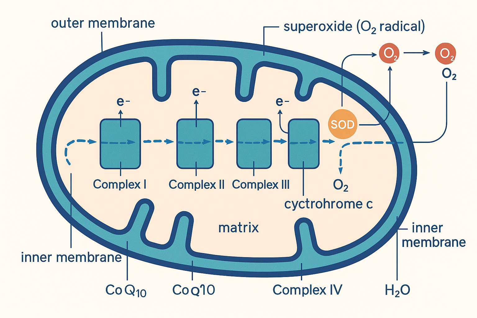 Where it begins: as electrons travel down the electron transport chain, a small fraction leak at Complexes I and III onto oxygen, forming superoxide. The mitochondrion is the brain's largest source of reactive oxygen species — and the first place defence must act.
Part 4 — The antioxidant defence system
Against this onslaught the brain runs a layered, cooperative defence — not a single antioxidant but an integrated network in which the members regenerate one another and hand danger down a chain until it is neutralised as harmless water. It has three tiers: the enzymes that catalytically destroy reactive species, the small-molecule antioxidants that absorb and pass on radical damage, and the master genetic programme (Nrf2) that scales the whole thing up or down. We take them in turn.
Tier 1 — The enzymatic defences
These are protein enzymes that destroy reactive species catalytically — meaning each enzyme molecule neutralises target molecules over and over, thousands of times per second, without being consumed. They are the front line, and they are arranged as an assembly line that walks superoxide all the way down to water.
-
Superoxide dismutase (SOD) — the first enzyme in the line, and the one whose discovery founded the field. It catalyses the dismutation of superoxide: it takes two superoxide molecules and converts them into one hydrogen peroxide and one oxygen. ("Dismutation" means a reaction where two identical molecules react with each other, one being oxidised and the other reduced.) There are versions inside the mitochondria (a manganese-dependent form, MnSOD/SOD2, sitting right where superoxide leaks) and in the cytosol (a copper-zinc form, SOD1). SOD does not eliminate the danger — it converts superoxide into hydrogen peroxide, which must then be dealt with by the next enzyme. SOD without downstream H₂O₂ disposal would simply move the problem along.
-
Catalase — converts hydrogen peroxide directly into water and oxygen, extremely fast. It is abundant in the liver but, as noted, comparatively sparse in the brain — which is precisely why the brain relies so heavily on the glutathione-based route instead.
-
Glutathione peroxidase (GPx) — the brain's main hydrogen-peroxide-disposal enzyme, and the reason glutathione is so central here. GPx uses two molecules of reduced glutathione (GSH) to convert hydrogen peroxide (or a lipid peroxide) into water (or a harmless alcohol), in the process joining the two glutathiones into one oxidised GSSG. Critically, GPx contains the trace element selenium at its active site — it is a selenoenzyme. This is the direct mechanistic reason selenium is an essential antioxidant nutrient: without selenium, you cannot build functional glutathione peroxidase, and the brain's primary peroxide defence falters.
-
Peroxiredoxins (Prx) and the thioredoxin system — a second, parallel peroxide-disposal network that has become recognised as quietly enormous in importance. Peroxiredoxins are extremely abundant and are now thought to handle a large share of the cell's everyday hydrogen peroxide, and to act as the actual sensors that pass the H₂O₂ signal on to other proteins (the signalling role in Part 5). They are kept in working order by thioredoxin, another small protein, which is in turn recharged by thioredoxin reductase using — once again — NADPH. Notice the recurring theme: every one of these systems ultimately draws its reducing power from NADPH.
Tier 2 — Glutathione: the master small-molecule antioxidant
If the brain has one central antioxidant, it is glutathione. It is the most abundant antioxidant inside cells (present at millimolar concentrations — very high for a regulatory molecule), and the GSH/GSSG ratio is the benchmark of redox health. It earns its own deep treatment.
What it is. Glutathione is a small molecule built from three amino acids: glutamate, glycine, and — the important one — cysteine. The whole antioxidant action lives on the cysteine, because cysteine carries a thiol group (a sulphur–hydrogen group, written –SH). That sulphur–hydrogen bond is the business end: it readily donates a hydrogen and an electron to neutralise a radical or to fuel glutathione peroxidase. When it does so, the sulphur is left with an unpaired electron, and it satisfies that by bonding to the sulphur of a second glutathione molecule — forming a disulphide bond (–S–S–) that links two glutathiones together. The linked pair is the oxidised form, GSSG.
The cycle. This is the elegant part, and it is genuinely a cycle, not a one-shot consumption:
- Two reduced glutathiones (GSH) hand over their electrons — either directly to a radical, or via glutathione peroxidase to destroy hydrogen peroxide — and in doing so are joined into one oxidised GSSG.
- An enzyme called glutathione reductase then regenerates the glutathione: it splits the disulphide bond and reduces GSSG back into two fresh GSH, ready to go again.
- The electrons for that regeneration step come from NADPH — closing the loop back to the cell's master reducing currency.
So glutathione is not used up like a fuel; it is cycled between reduced and oxidised forms, and the whole cycle is ultimately powered by NADPH (which the cell generates largely through a glucose-handling route called the pentose phosphate pathway). A cell "runs out" of glutathione protection only when production of reactive species overwhelms the regeneration capacity, or when raw materials run short.
flowchart LR
    H2O2["H2O2 / lipid peroxide<br/>(threat)"] -->|"glutathione peroxidase<br/>(needs selenium)"| H2O["H2O / harmless alcohol ✓"]
    GSH["2 × GSH<br/>(reduced · active)"] -->|donate electrons| GSSG["GSSG<br/>(oxidised · spent)"]
    H2O2 -.consumes.-> GSH
    GSSG -->|"glutathione reductase"| GSH
    NADPH["NADPH<br/>(reducing currency)"] -->|powers regeneration| GSSG
    CYS["Cysteine<br/>(rate-limiting raw material)<br/>← NAC / NACET supply this"] -.builds.-> GSH
The rate-limiting step — and why this matters for supplements. Of glutathione's three building blocks, glutamate and glycine are abundant, but cysteine is usually the limiting raw material — its availability sets the ceiling on how much glutathione a cell can make. This single fact is the mechanistic basis of an entire supplement category:
- NAC (N-acetylcysteine) is a stabilised, absorbable delivery form of cysteine. You do not take glutathione's building block "cysteine" directly because free cysteine is unstable and poorly absorbed; NAC is the workaround. By supplying the rate-limiting ingredient, NAC raises the cell's capacity to make glutathione — which is why it is used clinically (it is the antidote to paracetamol/acetaminophen overdose, precisely because that overdose kills the liver by exhausting glutathione, and NAC refills it).
- NACET (N-acetylcysteine ethyl ester) is a more lipophilic (fat-soluble) modification of NAC designed to cross membranes — and the blood–brain barrier — more readily, which is the rationale for its use as a more brain-penetrant glutathione precursor.
- Glutathione itself can be supplemented directly, though its oral absorption and ability to reach the brain intact are more contested than simply supplying cysteine and letting the cell build its own.
This is also a first hint at the calibration theme: supplying cysteine raises glutathione when cysteine is the bottleneck. If it is not the bottleneck — if the limit is selenium, or NADPH, or something else — then loading cysteine does less than the marketing implies.
Tier 3 — Small-molecule antioxidants beyond glutathione
Glutathione works mostly in the watery interior of the cell. Other small antioxidants cover other compartments, and they regenerate one another in a relay:
- Vitamin E — the chain-breaking antioxidant that lives inside membranes, where glutathione cannot reach, and stops the lipid-peroxidation cascade in the brain's vulnerable PUFA membranes. Its full mechanism (donate a hydrogen, become a harmless tocopheroxyl radical, get regenerated) is covered in its deep dive.
- Vitamin C (ascorbate) — the principal water-soluble antioxidant, and the molecule that regenerates spent vitamin E at the membrane surface, passing the absorbed damage back into the water phase where glutathione can handle it. The brain actively concentrates vitamin C to several times blood levels, underscoring its importance there.
- CoQ10 / ubiquinol — the same electron-shuttle from the electron transport chain doubles as a membrane antioxidant and also regenerates vitamin E, which is why the two are synergistic.
- Uric acid — not a supplement but worth knowing: it is one of the largest-capacity antioxidants in human blood plasma, accounting for a substantial fraction of plasma antioxidant power. (Humans, unusually among mammals, lost the enzyme that breaks uric acid down, and one hypothesis is that we kept high uric acid partly for its antioxidant value — a trade-off against its role in gout.)
- Melatonin — better known as the sleep hormone, but it is also a potent, lipophilic, brain-penetrant antioxidant that crosses into mitochondria, and several of its breakdown products are themselves antioxidants (a "cascade" of protection). This dual role is part of why melatonin is studied for neuroprotection well beyond sleep.
The picture to hold is a relay: a radical that attacks a membrane fat is intercepted by vitamin E; vitamin E is regenerated by vitamin C and CoQ10; the damage passes into the water phase and is absorbed by glutathione; glutathione is regenerated by NADPH. No single antioxidant works alone, which is the deepest reason why mega-dosing one isolated antioxidant so often disappoints — you can saturate one link while the chain is bottlenecked elsewhere.
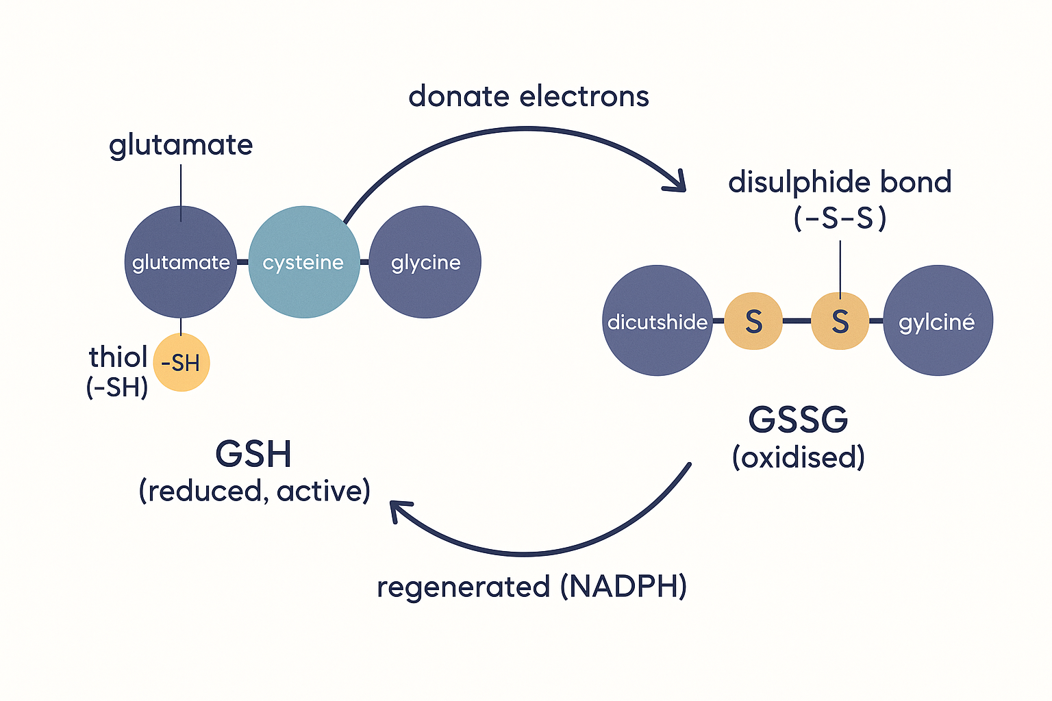 The cell's master redox couple: reduced glutathione (GSH) carries a reactive sulphur–hydrogen thiol on its cysteine; after neutralising a threat, two glutathiones join at a sulphur–sulphur bond to form oxidised GSSG. The GSH/GSSG ratio is the standard readout of a cell's redox health.
The master switch — the Nrf2 / ARE programme
All of the above — SOD, glutathione peroxidase, the enzymes that make glutathione, the thioredoxin system — are proteins, and proteins are made by switching on genes. The brain does not run its antioxidant defence at a fixed level; it scales it up when it senses oxidative threat, through a single master transcription factor: Nrf2.
The mechanism is itself a beautiful piece of redox sensing:
- Nrf2 (full name nuclear factor erythroid 2–related factor 2) is a protein that, when it reaches the cell nucleus, switches on a whole battery of antioxidant and detoxification genes — those genes share a common DNA tag in their control region called the Antioxidant Response Element (ARE). Switch on Nrf2 and you turn up the production of glutathione-synthesis enzymes, glutathione peroxidase and reductase, thioredoxin, NADPH-generating enzymes, and more, all at once.
- Under calm conditions, Nrf2 is held captive in the cytoplasm by a partner protein called Keap1, which constantly tags Nrf2 for destruction — keeping the antioxidant programme idling.
- Keap1 is itself a redox sensor. It is studded with reactive cysteine thiols (the same –SH chemistry as glutathione). When oxidants or certain reactive plant chemicals modify those thiols, Keap1 changes shape and releases Nrf2. Freed Nrf2 travels to the nucleus and fires up the whole ARE gene programme. In other words: the cell detects oxidative stress and responds by manufacturing more of its own defences.
This is profoundly important for how interventions actually work. Many of the most robust "antioxidant" compounds — including the sulforaphane in broccoli sprouts and curcumin — do not act mainly by mopping up radicals themselves. They are mild Keap1 irritants: they poke the sensor, release Nrf2, and trigger the cell to build its own, far larger and self-renewing antioxidant response. This is a hormetic mechanism — a small, controlled stress that provokes an adaptive over-response — and it is mechanistically superior to swallowing a direct antioxidant, because an enzyme system the cell builds for itself is catalytic, regenerating, and correctly localised, whereas a swallowed antioxidant is consumed once. Hold onto this distinction; it is the hinge of the calibration argument in Part 6.
flowchart TD
    CALM["Calm cell"] --> KEAP["Keap1 holds Nrf2 captive<br/>+ tags it for destruction"]
    KEAP --> IDLE["Antioxidant genes idling"]
    STRESS["Oxidants OR<br/>sulforaphane / curcumin"] -->|"modify Keap1's<br/>reactive thiols"| RELEASE["Keap1 releases Nrf2"]
    RELEASE --> NUC["Nrf2 enters nucleus,<br/>binds ARE"]
    NUC --> GENES["Turns ON defence genes:<br/>glutathione synthesis · GPx ·<br/>thioredoxin · NADPH supply"]
    GENES --> DEFENCE["Cell builds its OWN<br/>larger, self-renewing defence"]
Part 5 — Redox as a signal, not just damage — and the hormetic U-curve
Here is where the popular understanding of this topic breaks down, and where the genuinely sophisticated picture begins. Reactive oxygen species are not only damaging by-products to be eliminated. At low, controlled concentrations they are essential signalling molecules that the brain cannot function without. A cell scrubbed completely free of reactive oxygen would not be healthy — it would be deaf to a whole channel of information and unable to adapt. This is the single most important idea on the page, and the one most at odds with "antioxidants = always good."
Hydrogen peroxide as a deliberate messenger
Recall that hydrogen peroxide is mild, stable, and membrane-permeable — exactly the properties you would want in a signalling molecule. The cell exploits this. A controlled puff of H₂O₂ (made deliberately by NOX enzymes, or arising from mitochondrial activity) acts as a message by reversibly oxidising specific cysteine thiols on target proteins — flipping them between an "off" and "on" shape, just like the more familiar on/off switching by phosphate groups. Because the modification is reversible (the thioredoxin and glutathione systems can switch it back off), it works as a genuine, tunable signal rather than damage. This redox signalling regulates:
- Growth-factor and insulin signalling — many growth and metabolic signals deliberately generate a local burst of H₂O₂ as part of their normal action; the burst transiently inhibits the "off-switch" enzymes that would otherwise terminate the signal. This is one route by which intranasal insulin (below) and redox tone influence brain signalling.
- The Nrf2 response itself — as we just saw, oxidants are the trigger for building more defence.
- Adaptation to exercise and metabolic stress — the reactive-oxygen burst from exercising mitochondria is a large part of the signal that tells cells to build more mitochondria and more antioxidant enzymes. This is why high-dose antioxidants taken around exercise can blunt some of training's beneficial adaptations — you mop up the very signal that drives the adaptation.
Redox modulation of neuronal plasticity
In the brain specifically, reactive species are woven directly into learning and memory. The NMDA receptor — the central molecular switch for synaptic plasticity (the strengthening of connections that underlies learning, especially a process called long-term potentiation, LTP) — is redox-sensitive: it carries cysteine thiols that, depending on their oxidation state, tune the receptor's activity up or down. More broadly, a modest level of reactive oxygen (much of it from NOX enzymes activated during neuronal firing) is required for normal LTP — experimentally, scavenging reactive oxygen too aggressively impairs memory formation. The reactive species the brain produces during intense activity are part of the mechanism by which that activity reshapes the brain.
So reactive oxygen in the brain is genuinely double-edged: the same chemistry that, uncontrolled, destroys neurons is, controlled, the medium of adaptation, signalling, and memory.
The hormetic U-curve — the central calibration principle
This sets up the most important conceptual tool on the page: the relationship between reactive-oxygen level and neuronal function is not a downward slope ("more oxidation, always worse") but a U-shaped (or inverted-U) curve.
- Too little reactive oxygen — the over-scavenged, "reductive stress" state — and signalling fails: blunted plasticity, impaired adaptation, sluggish Nrf2 defences, and even a paradoxical vulnerability because the cell never trained its defences. (Yes, there is such a thing as reductive stress — an excess of reducing power that is itself pathological; it is less famous than oxidative stress but real.)
- A moderate, dynamic level — the healthy window — and reactive oxygen does its signalling job, drives adaptation, and is comfortably handled by defences kept appropriately exercised. This is the optimum.
- Too much — true oxidative stress — and the destruction described throughout this page takes over: lipid peroxidation, protein and DNA damage, cell death.
The goal is therefore not to drive reactive oxygen as low as possible. The goal is to sit in the middle of the U. This single reframing dissolves the antioxidant paradox: the reason huge clinical trials of high-dose vitamin E or beta-carotene showed no benefit or slight harm is that, in people who were not actually oxidatively stressed, the supplements pushed them down the wrong side of the curve — into blunted signalling and impaired adaptation. Antioxidants help the person on the right-hand (over-oxidised) side of the curve; they can harm the person already near the optimum by shoving them left.
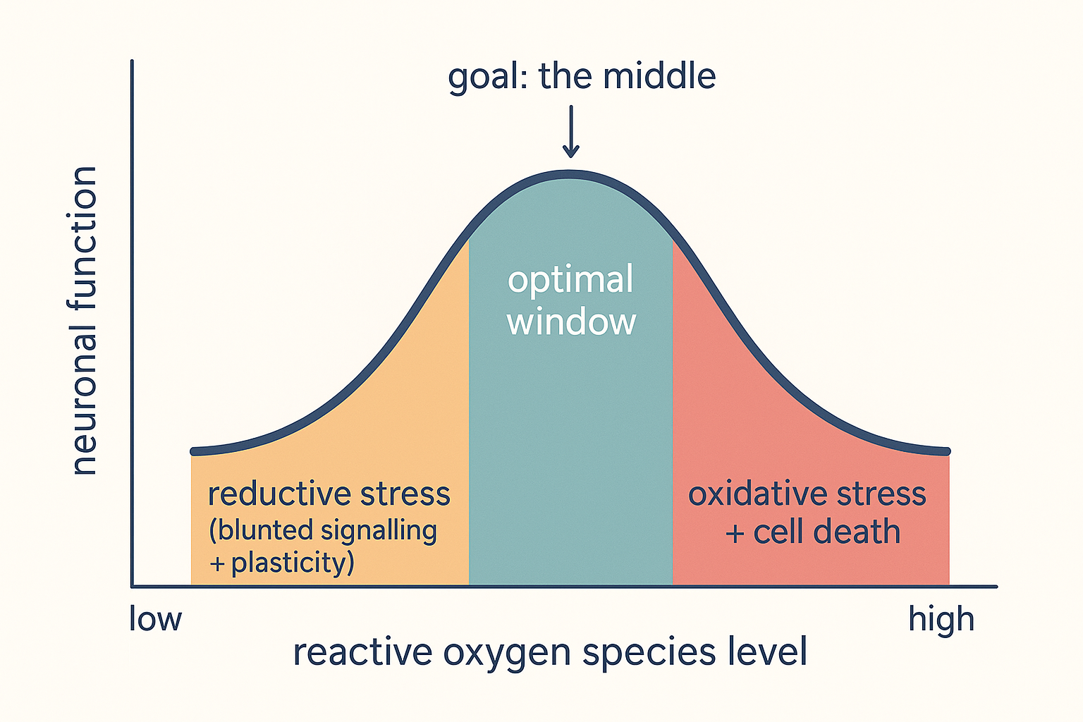 The calibration principle in one curve: neuronal function peaks at a moderate level of reactive oxygen, not at zero. Too little (reductive stress, blunted signalling and plasticity) is as harmful as too much (oxidative stress, lipid peroxidation, cell death). The aim of any intervention is to move toward the middle — which means antioxidants help only those who start on the right.
Part 6 — When the balance breaks: oxidative stress and disease
When production chronically outstrips defence, the brain tips off the right edge of the U-curve into sustained oxidative stress, and the consequences are central to the major neurological diseases. This section is also the bridge to the companion Neurotoxicity & Neuroprotection page.
Lipid peroxidation — the membrane catastrophe. The brain's PUFA-rich membranes are the prime target, and the Vitamin E deep dive describes the mechanism in full. A radical steals a hydrogen from a membrane PUFA, turning it into a radical that attacks the next fat — a chain reaction that rips through the membrane, converting structural fat into toxic, reactive breakdown products, the best-known being 4-HNE (4-hydroxynonenal) and MDA (malondialdehyde). These are not inert waste: they are themselves reactive, drift to other proteins and cripple them, and are used in the lab as the standard markers of oxidative damage. In the brain this degrades synaptic membranes, myelin, and the blood–brain barrier itself, and — when iron-driven and unchecked by glutathione peroxidase — culminates in the iron-dependent cell death called ferroptosis.
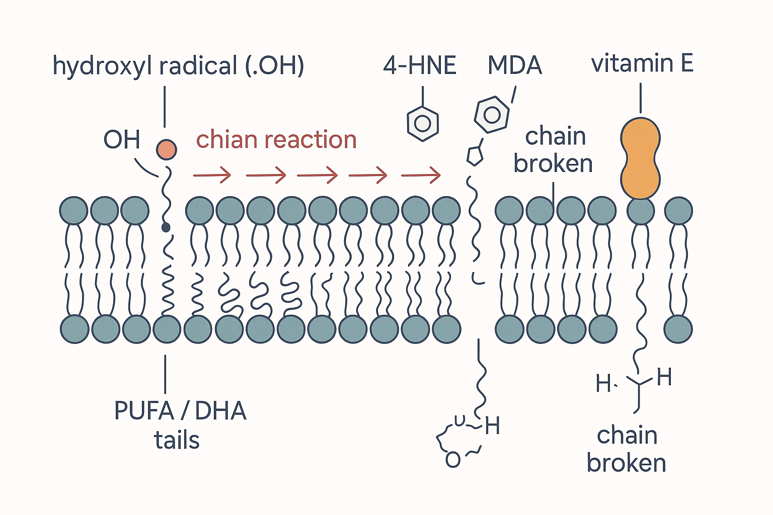 The membrane chain reaction in a neuron: a single radical strips a hydrogen from a polyunsaturated DHA tail, and the damage propagates fat-to-fat through the synaptic membrane — spawning toxic 4-HNE and MDA — until a vitamin E molecule embedded among the fats donates a hydrogen and breaks the chain. The brain's DHA-rich membranes make this its signature mode of oxidative damage.
Neurodegeneration. Oxidative stress is a consistent, early feature of the major neurodegenerative diseases, both as cause and accelerant:
- In Parkinson's disease, the neurons that die are the dopamine neurons of the substantia nigra — a region that is unusually exposed on every redox axis at once: it is iron-rich, and the metabolism of dopamine itself generates hydrogen peroxide and reactive quinones as by-products. Dopamine neurons are, in effect, living in a chemically hostile microclimate; mitochondrial Complex I dysfunction and glutathione depletion are among the earliest detectable changes. This is also why neuroprotection is such a recurring theme among people using dopaminergic stimulants — the dopamine system carries an intrinsic oxidative burden.
- In Alzheimer's disease, oxidative damage to lipids, proteins, and DNA appears early, and there is a vicious loop with the amyloid-beta protein: amyloid both generates reactive oxygen (partly via bound metal ions) and is produced more under oxidative stress, each feeding the other.
Ischaemia–reperfusion injury — the paradox of restored blood flow. This is one of the clearest demonstrations that oxidative stress is a distinct, mechanistic thing. In a stroke (or any episode where blood flow is cut off and then restored), a large part of the damage occurs not during the oxygen deprivation but during the re-oxygenation — when blood flow returns. Starved mitochondria, when suddenly re-supplied with oxygen, produce a massive burst of superoxide; combined with the nitric oxide present, this generates a flood of peroxynitrite, and the antioxidant defences (already depleted by the crisis) are overwhelmed. The tissue is, paradoxically, damaged by the return of oxygen. This is why antioxidant and reactive-oxygen-targeted strategies are a major research focus for stroke and traumatic brain injury — and why compounds marketed for "stroke recovery" and "neuroprotection under hypoxia" (such as mexidol, widely used in Russia for exactly this indication) are framed around blunting this reperfusion burst.
The inflammation loop. Oxidative stress and inflammation are not separate problems but a self-reinforcing cycle. Reactive oxygen activates the master inflammatory switch NF-κB; the resulting inflammation activates microglia, whose NOX enzymes produce more reactive oxygen; lipid-peroxidation products are themselves inflammatory triggers. In the brain this loop is called neuroinflammation, and breaking it at the redox step is one rationale for antioxidant neuroprotection. Each page's villain is the other's accomplice.
Part 7 — The bioenergetic angle, and the database tie-in
The Ray-Peat-influenced, bioenergetic framing that runs through this site approaches brain redox from a characteristic angle, and it is worth presenting clearly — then calibrating honestly against the mainstream.
The bioenergetic thesis. In this view, the root problem is rarely "too few antioxidants" and almost always a failing energy metabolism. The logic: a mitochondrion running cleanly and efficiently — with electrons flowing briskly all the way to oxygen and out as water — leaks fewer radicals in the first place. A mitochondrion that is sluggish, "backed up," or poisoned (by, in this framework, an excess of polyunsaturated fat in its membranes, by low thyroid, by endotoxin) leaves electrons sitting on the carriers, where they leak onto oxygen as superoxide. So the bioenergetic priority is to fix the energy metabolism — keep the electron flow brisk and complete — rather than to chase radicals after the fact. Concretely this means the familiar Peat levers: avoid PUFA (fewer oxidation-prone fats in the membranes means less lipid peroxidation to defend against — the same logic as the vitamin E page), support thyroid and a brisk metabolic rate, value CO₂ and a well-oxygenated, efficient respiration, and favour antioxidants that are also metabolic supports.
Methylene blue as the emblematic compound. Methylene blue is the bioenergetic world's favourite redox tool precisely because it is a redox molecule. At low doses it acts as an alternative electron carrier — it can pick up electrons within the mitochondria and ferry them onward, in effect providing a "bypass" around a partially blocked electron transport chain. By keeping electrons moving rather than stagnating, it is argued to reduce the leak that produces superoxide, while supporting ATP output. It is the clearest example of the bioenergetic preference for improving electron flow over scavenging the products of poor flow — a redox cycler rather than a sacrificial antioxidant. (Calibration: methylene blue's low-dose mitochondrial effects are real and studied, but it is hormetic and dose-sensitive — at higher doses it generates reactive oxygen and inhibits an enzyme that matters greatly for anyone on serotonergic drugs, so it is not a "more is better" compound.)
The database tie-in: "improving redox balance" as a dopaminergic mechanism
This page connects directly to a stack circulating in the community. The user @aestheticprimal, describing a "dopamine enhancement stack," makes a striking and specific mechanistic claim — that the benefit of two of his core agents is mediated through redox:
"Cerebrolysin seems to enhance Dopaminergic transmission mainly through improving redox balance (total antioxidant capacity)..."
and, of the second agent:
"Intranasal Insulin... Seems to be mainly mediated through improving redox balance in the brain."
This is exactly the framework this page has built, applied in the wild — and it is worth dissecting with the calibration the rest of the page demands.
- The claim rests on the genuine biology of Part 5: dopamine signalling is redox-sensitive, and the dopamine neurons of the substantia nigra (Part 6) live under an intrinsic oxidative burden because dopamine metabolism itself generates hydrogen peroxide and reactive quinones. A treatment that genuinely improved the redox tone of those neurons — raising "total antioxidant capacity" — could plausibly support their health and firing, which is a coherent (if hard-to-prove) route to "a much higher dopamine baseline."
- Intranasal insulin is the more mechanistically grounded of the two. Insulin signalling in the brain recruits exactly the redox-coupled pathways of Part 5 (insulin signalling deliberately uses local H₂O₂; it also supports mitochondrial function and Nrf2-linked defences), and the intranasal route is designed to deliver it to the brain while bypassing systemic blood sugar effects. There is, as the author notes, a genuinely large literature on intranasal insulin and cognition — though framing its entire benefit as "redox balance" is a simplification of a broader metabolic action.
- Cerebrolysin (a mixture of neuropeptides) has its principal evidence in neurotrophic and neuroprotective effects; "improving redox balance" is one proposed contributor among several (the author himself adds GDNF-receptor activation), and the dopaminergic-specific claim is, by his own admission, more personal experience than established trial data.
The honest reading: the redox framing is mechanistically literate and points at real biology, and it correctly identifies the dopamine system as redox-vulnerable. But "improving redox balance / total antioxidant capacity" is doing a lot of work as a catch-all explanation, and — per the hormetic U-curve of Part 5 — "more antioxidant capacity" is only beneficial if you start on the over-oxidised side of the curve. The neuroprotection-stacking instinct that runs through this community (pairing stimulants with mexidol, NACET, and similar) is well-founded to the extent that dopaminergic stimulation genuinely raises the oxidative load on vulnerable neurons — which it does. The error to avoid is treating antioxidant capacity as a free good to be maximised, rather than a balance to be calibrated.
Putting it all together
Step back and view the whole system:
-
Redox is electron bookkeeping. Oxidation is electron loss, reduction is electron gain, and they are always paired. An "antioxidant" is just a molecule willing to donate electrons. The cell's redox state is read off three couples — NAD⁺/NADH (energy), NADPH/NADP⁺ (the reducing currency for defence), and GSH/GSSG (the master antioxidant readout). Oxidative stress is a balance concept: production of reactive species exceeding the capacity to neutralise them.
-
The brain is the worst case on every axis at once: highest oxygen throughput (most radical production), most oxidation-prone PUFA membranes, catalytic iron, modest enzymatic defence (low catalase — hence its reliance on glutathione), and irreplaceable post-mitotic neurons that accumulate damage permanently.
-
The reactive species form a cascade: electron leak at ETC Complexes I and III (and deliberate NOX enzymes) makes superoxide → SOD converts it to hydrogen peroxide (mild, stable, and the brain's main signal) → which is either safely removed to water by glutathione peroxidase/catalase/peroxiredoxins, or, with free iron, becomes the indiscriminate hydroxyl radical. Separately, nitric oxide plus superoxide makes peroxynitrite.
-
Defence is a three-tier network: enzymes (SOD → glutathione peroxidase/catalase/peroxiredoxins) that catalytically destroy species; small-molecule antioxidants in a regenerating relay (vitamin E in membranes, regenerated by vitamin C and CoQ10; glutathione in the cytosol, regenerated by NADPH); and the master gene programme Nrf2/ARE, which the cell switches on to build more of its own defence when it senses oxidant stress. Glutathione is central, its synthesis limited by cysteine — which is why NAC and NACET work — and its disposal enzyme requires selenium.
-
Reactive oxygen is also a signal, not only damage: H₂O₂ tunes growth, insulin, and Nrf2 signalling, and a moderate level is required for NMDA-receptor-dependent plasticity and memory. Hence the hormetic U-curve: function peaks at a moderate level, with both too little (reductive stress, blunted adaptation) and too much (oxidative stress, cell death) being harmful. The goal is the middle of the curve, not zero — which is exactly why indiscriminate high-dose antioxidants fail or harm.
-
When the balance breaks, the brain tips into sustained oxidative stress: lipid peroxidation (4-HNE, MDA, ferroptosis), the early oxidative signature of Parkinson's (iron- and dopamine-driven) and Alzheimer's (the amyloid loop), the re-oxygenation burst of stroke, and the self-reinforcing oxidative-inflammatory loop of neuroinflammation.
-
The bioenergetic framing prioritises fixing the energy metabolism so fewer radicals leak in the first place (lower PUFA, support thyroid, keep electron flow brisk) over scavenging them afterward — with methylene blue as the emblematic redox-cycling tool. The community's "improving redox balance" framing for dopaminergic agents (cerebrolysin, intranasal insulin) is mechanistically literate and points at the genuine redox-vulnerability of dopamine neurons — but must be held to the same calibration: antioxidant capacity is a balance to optimise, not a quantity to maximise.
The single unifying idea: the brain does not want to eliminate oxidation — it wants to govern it. Reactive oxygen is simultaneously the brain's most dangerous by-product and one of its essential signals. Health is not the absence of oxidation but its precise regulation — enough to signal and adapt, never so much as to destroy. Every intervention on this page should be judged by whether it moves a given brain toward that balanced middle, not simply down.
Related Compounds & Deep Dives
The glutathione system (the brain's central antioxidant)
- NAC (N-acetylcysteine) — delivers cysteine, the rate-limiting raw material for glutathione synthesis; the clinical glutathione-replenisher.
- NACET — a more lipophilic, brain-penetrant cysteine donor; the "superior NAC" framing rests on better blood–brain-barrier crossing.
- Glutathione — the master antioxidant tripeptide itself; the GSH/GSSG ratio is the standard readout of redox health.
- Selenium — the essential cofactor at the active site of glutathione peroxidase, the brain's main hydrogen-peroxide-disposal enzyme.
The membrane antioxidant relay
- Vitamin E deep dive — the chain-breaking, membrane-resident antioxidant that stops lipid peroxidation in the brain's PUFA membranes; full mechanism and the alpha-vs-gamma debate.
- Vitamin C — the principal water-soluble antioxidant; regenerates spent vitamin E; actively concentrated by the brain.
- CoQ10 / ubiquinone and ubiquinol — the electron-transport-chain shuttle that doubles as a membrane antioxidant and regenerates vitamin E.
- Melatonin — beyond sleep, a brain- and mitochondria-penetrant antioxidant with antioxidant breakdown products.
Mitochondrial / redox-cycling tools
- Methylene blue — alternative mitochondrial electron carrier; reduces electron leak by keeping flow moving rather than scavenging products (dose-sensitive and hormetic).
- Mexidol (emoxypine succinate) — a brain-penetrant antioxidant used (in Russia) for stroke, hypoxia, and stimulant neuroprotection — i.e. the ischaemia-reperfusion oxidative burst of Part 6.
The bioenergetic / dopaminergic stack (database tie-in)
- Cerebrolysin — neuropeptide mixture argued to enhance dopaminergic transmission partly "through improving redox balance (total antioxidant capacity)."
- Intranasal insulin — brain-targeted insulin whose cognitive benefit is framed as "mainly mediated through improving redox balance in the brain"; engages the redox-coupled insulin-signalling biology of Part 5.
Related foundations
- Cellular Energy: From Fuel to ATP — the electron transport chain whose electron leak is the brain's primary reactive-oxygen source.
- Inflammation — the self-reinforcing oxidative–inflammatory loop; antioxidant defence and resolution overlap heavily.