Brain Waves (Neural Oscillations)
Why the brain hums at a handful of fixed rhythms, how a few million cells whispering in time produce a signal you can read off the scalp, and what each band — delta, theta, alpha, beta, gamma — actually does for sleep, memory, attention, and consciousness itself. Plus an honest look at neurofeedback, binaural beats, and the 40 Hz gamma research that everyone is excited about.
In 1924 a reclusive German psychiatrist named Hans Berger taped electrodes to the scalp of his son, connected them to a galvanometer, and recorded a faint rhythmic voltage rising and falling roughly ten times a second. He had captured the first human electroencephalogram (EEG) — literally "electric-brain-writing" — and the dominant rhythm he saw came to be called the alpha wave. Berger noticed something strange: the moment his subject opened their eyes or began to think, the steady ten-per-second rhythm vanished. The brain, it turned out, was not silent at rest; it was oscillating, and the oscillation changed with mental state. He published in 1929 to widespread scepticism — colleagues assumed he was recording electrical artefact, not the brain — until others reproduced it.
The second landmark came in sleep. In 1953 Eugene Aserinsky and Nathaniel Kleitman discovered that sleep is not a uniform descent into oblivion but a cycle, punctuated by periods of darting eye movements and a strangely wakeful-looking EEG — REM sleep, the stage of vivid dreaming. In 1957 William Dement and Kleitman formally mapped the night into EEG-defined stages, showing that distinct brain rhythms label distinct states of consciousness. Brain waves became the alphabet with which we read the sleeping and waking mind.
A century on, oscillations are no longer just a readout — they are increasingly understood as a mechanism. Current work treats rhythms as the brain's way of routing information (communication-through-coherence), packaging memories (theta–gamma coupling), and even as a therapeutic target: driving the brain at 40 Hz with flickering light and sound is in phase-III trials for Alzheimer's disease. This page builds the whole picture from first principles.
Why this page exists
Almost everything in the biohacking world that touches the brain eventually invokes "brain waves" — meditation apps promising "more alpha," binaural-beat tracks claiming to push you into "theta for deep relaxation," neurofeedback clinics training "SMR for focus," sleep trackers reporting your "deep-sleep delta." Most of this language is used loosely, and some of it is marketing nonsense draped over a real and beautiful piece of neuroscience.
The real thing is worth understanding properly, because it is the temporal dimension of everything the neuroscience of cognition page described. That page explained which chemicals carry signals and which circuits they run in — the spatial wiring. This page explains the timing: how populations of neurons coordinate when they fire, why that coordination matters as much as the wiring, and how the brain uses rhythm to bind, route, gate, and store information. By the end you should be able to read any brain-wave claim — a neurofeedback protocol, a binaural track, a sleep-stage readout, the 40 Hz gamma hype — and say what is mechanistically plausible, what is established, and what is wishful.
We build it in order: what an oscillation physically is, how the brain generates one, the bands one by one, what rhythms do, how we measure them, and finally the states and applications.
What a neural oscillation actually is
Start with a single neuron. From the cognition page, recall that a neuron is a nerve cell, and that it signals by firing an action potential — a brief, all-or-nothing electrical spike that races down its output fibre (the axon) and triggers the release of neurotransmitter onto the next cell. The spike itself is the neuron's output.
But a neuron also has inputs — thousands of them, arriving on its branching receiving structures (the dendrites). Each incoming signal nudges the cell's internal voltage slightly up or slightly down. An excitatory input (mostly glutamate) makes the inside of the cell a little less negative, pushing it toward firing — an excitatory postsynaptic potential (EPSP). An inhibitory input (mostly GABA, gamma-aminobutyric acid) makes the inside more negative, pushing it away from firing — an inhibitory postsynaptic potential (IPSP). These postsynaptic potentials are small, graded, and relatively slow — they last tens of milliseconds, far longer than the sub-millisecond spike.
Here is the crucial point that the whole page rests on: EEG does not measure spikes. It measures the summed postsynaptic potentials of large populations of neurons.
Why? Two reasons. First, geometry. The cortex's principal cells, the pyramidal neurons, are arranged like trees in an orchard — all aligned in parallel, perpendicular to the cortical surface, with a long shaft (the apical dendrite) reaching up toward the surface and the cell body sitting deeper. When such a cell receives excitatory input near the top of its dendrite, positive ions flow into the cell there, leaving the surrounding fluid slightly negative at the top and slightly positive deeper down. The cell briefly becomes a tiny electrical dipole — a little battery with a plus and a minus end. Because all the pyramidal cells point the same way, their dipoles line up and add together instead of cancelling. This aligned, "open-field" geometry is what makes the cortex electrically visible from outside the head at all.
Second, timing. A single cell's dipole is far too feeble to detect through skull and scalp. To produce a measurable voltage, millions of these dipoles must point the same way at the same time. And that is the entire reason synchrony matters: a voltage you can read on the scalp exists only when a large population of neurons is doing the same thing in rhythm. The summed, smoothed electrical field inside the tissue is called the local field potential (LFP); the EEG is essentially that field, attenuated and blurred, read at the scalp.
This explains a deep and counter-intuitive fact. A bigger EEG wave does not mean more brain activity — it often means less, but more synchronised. When a million cells idle in lock-step, their dipoles sum into a large, slow wave (this is what deep sleep looks like). When the same cells are all busy doing different things — actively computing — their dipoles point every which way and largely cancel, leaving a small, fast, "desynchronised" trace. High-amplitude EEG is the signature of idling in unison; low-amplitude fast EEG is the signature of active, independent processing. Berger's alpha rhythm collapsing the instant his subject opened their eyes is exactly this: synchronous idling giving way to busy, desynchronised work.
So a neural oscillation is a rhythmic rise and fall in this summed field, produced by a population of neurons whose excitability — and therefore their tendency to fire — waxes and wanes together in a repeating cycle. The frequency of that cycle (how many times per second the field swings up and down, measured in hertz, Hz) is what defines the "band." The question now is: what makes a population cycle in the first place?
How a rhythm is generated: the excitation–inhibition loop
Rhythms do not require a metronome. They emerge, almost inevitably, from one structural fact: the cortex is built from excitatory cells that drive each other and inhibitory cells that restrain them, wired into a loop. Any loop with a delay and a brake tends to oscillate — the same reason a thermostat overshoots, a crowd's applause synchronises, or feedback howls through a microphone.
The brake is supplied by a special class of GABAergic inhibitory interneurons — local cells that release GABA onto their neighbours. The single most important class for fast rhythms is the parvalbumin-expressing (PV) fast-spiking interneuron (named for a calcium-binding protein, parvalbumin, that they contain). These cells are extraordinary: they fire faster than any other neuron in the cortex (hundreds of spikes per second without fatiguing), they wrap their inhibitory terminals tightly around the cell bodies of dozens of nearby pyramidal neurons — the most powerful place to apply a brake — and a single PV cell can silence a whole local population in one synchronised stroke. They are the conductors that keep the orchestra in time.
The basic rhythm-generating loop works like this:
- A pool of excitatory pyramidal neurons fires, releasing glutamate.
- That glutamate excites the local PV interneurons, which fire a fraction of a millisecond later.
- The PV cells release GABA back onto the whole pyramidal pool at once, silencing it.
- The inhibition decays over a few milliseconds; as it fades, the pyramidal cells recover and fire again.
- Back to step 1.
Each loop takes a fixed amount of time, set largely by how long the GABA inhibition lasts — and that fixed loop-time is the period of the oscillation. Fast PV inhibition (decaying in ~25 milliseconds) produces a ~40-per-second rhythm: gamma. Slower inhibition produces slower bands. Because the inhibition resets the entire population simultaneously, every cell starts its next cycle together — the population is forced into synchrony.
flowchart LR
    PYR["Pyramidal cells fire<br/>(glutamate)"] -->|excite| PV["PV interneurons fire<br/>(GABA)"]
    PV -->|inhibit whole pool| SIL["Pyramidal pool silenced"]
    SIL -->|GABA decays<br/>over ms| REC["Cells recover"]
    REC -->|fire together| PYR
Neuroscientists distinguish two flavours of this engine. When the excitatory cells lead and recruit the inhibition (the loop above), it is called PING — pyramidal–interneuron network gamma. When the inhibitory cells are driven hard enough to synchronise among themselves through mutual inhibition, pacing the network almost on their own, it is called ING — interneuron network gamma. They are not rivals; the network slides between them depending on how strong the excitatory drive is. The headline is the same either way: the rhythm is set by the inhibitory interneurons, not the excitatory cells. The brain's clocks are made of brakes. This is why the GABA system — the subject of its own page — sits at the heart of every fast oscillation, and why drugs that prolong GABA inhibition (benzodiazepines, alcohol) visibly slow and reshape the EEG.
How a rhythm is generated: the thalamocortical loop
The local E/I loop explains fast, local rhythms. But the slowest, most global rhythms — the alpha of relaxed wakefulness, the spindles and slow waves of sleep — are paced by a second, larger loop running between the cortex and a deep structure beneath it: the thalamus.
The thalamus is the brain's central relay — almost every sensory stream (sight, sound, touch) passes through it on the way to the cortex, and the cortex sends massive feedback projections back down. Wrapped around the thalamus like a shell is a thin sheet of purely inhibitory (GABAergic) cells, the thalamic reticular nucleus (TRN). This three-part circuit — cortex, thalamic relay cells, and the inhibitory TRN — forms a resonant loop with its own characteristic rhythms.
The key to the loop is a special property of thalamic relay neurons: they have a voltage-dependent calcium channel (the "T-type" channel) that gives them two modes of firing. When the cell is at a normal resting voltage it relays signals faithfully, spike for spike (tonic mode — the waking relay). But when the cell is hyperpolarised — pushed to a more negative voltage by a burst of inhibition from the TRN — that calcium channel de-inactivates, and when the inhibition lifts, the channel opens and the cell fires a rebound burst of spikes (burst mode). This rebound is the engine of the loop:
- The inhibitory TRN fires, hyperpolarising the thalamic relay cells.
- The inhibition lifts; the relay cells rebound-burst.
- That burst drives the cortex and loops back to re-excite the TRN.
- The TRN fires again, and the cycle repeats.
flowchart LR
TRN["Reticular nucleus<br/>(inhibitory)"] -->|inhibit| REL["Thalamic relay cells<br/>hyperpolarise"]
REL -->|inhibition lifts| BURST["Rebound burst<br/>(T-type Ca channel)"]
BURST -->|drive| CTX["Cortex"]
BURST -->|re-excite| TRN
CTX -->|feedback| TRN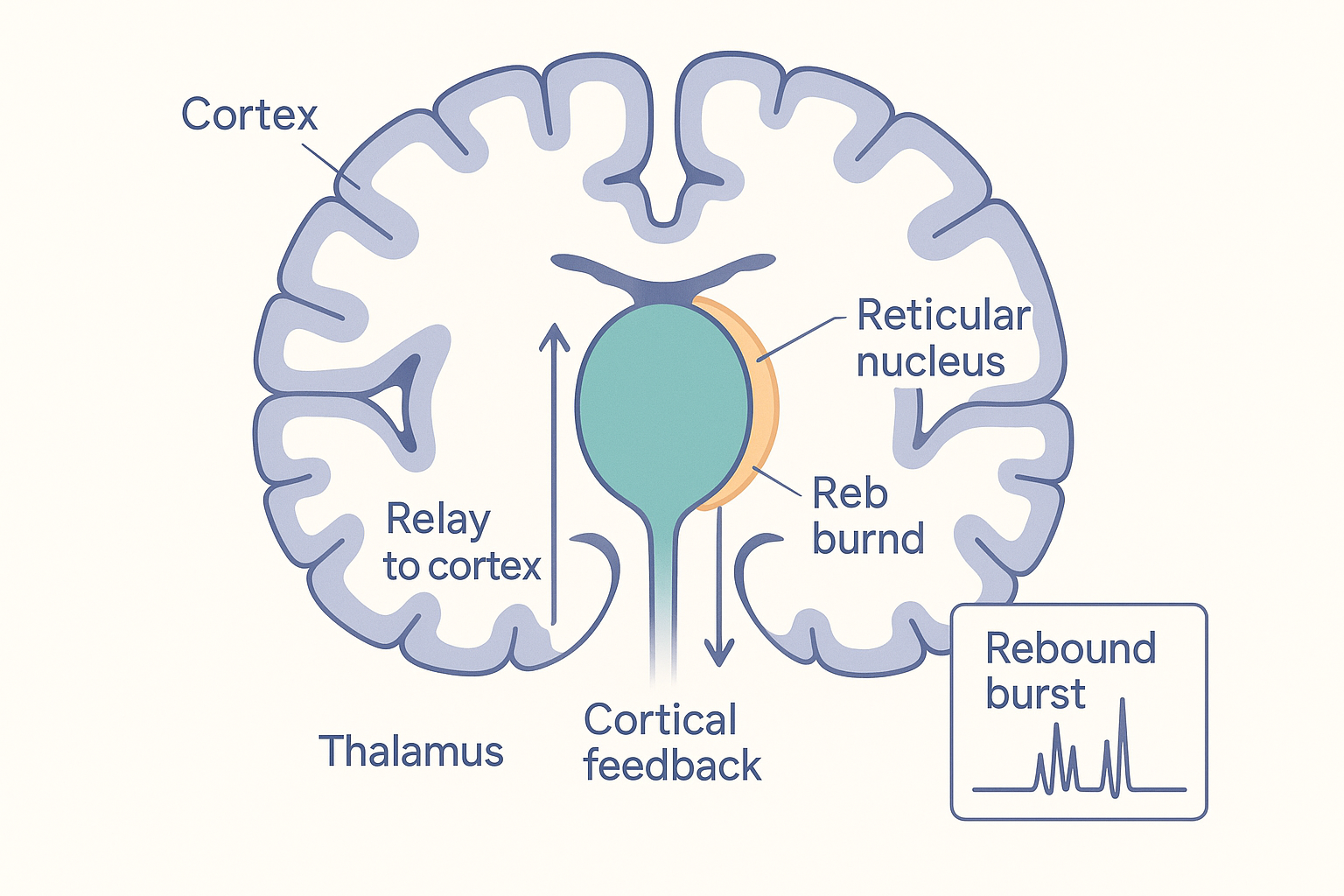 The anatomy behind the loop: the inhibitory reticular nucleus (a thin shell) wraps the thalamus deep in the brain, and reciprocal fibres connect it to the layered cortex above. The relay cells' rebound bursting — set by how deeply the reticular shell hyperpolarises them — paces alpha in waking and slows into spindles and delta as you fall asleep.
The speed of this loop depends on how deeply the relay cells are hyperpolarised, which in turn depends on the overall state of arousal — and that is set by neuromodulators rising up from the brainstem (acetylcholine, noradrenaline, serotonin, histamine; many introduced on the cognition and ANS pages). In relaxed wakefulness the loop produces alpha. As you fall asleep and the modulators withdraw, the relay cells hyperpolarise further, and the loop first produces sleep spindles, then slows all the way down into the great delta slow waves of deep sleep. This single loop, re-tuned by arousal chemistry, generates the entire descending sequence of sleep rhythms — the deep mechanistic reason that brain waves track states of consciousness, and the link to the sleep architecture page.
The bands, one by one
By long convention the EEG spectrum is divided into named frequency bands. The boundaries are somewhat arbitrary (different labs use slightly different cut-offs), and the bands are not separate "things" the brain emits so much as a continuum we have carved into ranges. But each range is dominated by particular generators and accompanies particular states, and the names are the lingua franca of the whole field. There is a beautiful inverse relationship running through them: the slower the rhythm, the larger its amplitude and the more brain it spans; the faster the rhythm, the smaller and more local it is. Slow waves are the whole orchestra playing one note; fast waves are small sections trading rapid phrases.
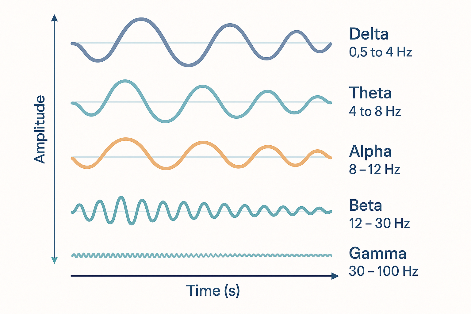 The five classical bands drawn to scale on a common timeline. Note the inverse trade-off: delta is slow and high-amplitude (synchronous idling of huge populations); gamma is fast and low-amplitude (small assemblies doing rapid, local work). Frequency rises and amplitude falls as you move down the stack.
Delta — 0.5 to 4 Hz — the deep-sleep slow wave
Delta is the slowest and tallest band: huge, slow swings of the field, up to 200 microvolts. It dominates the deepest stage of dreamless sleep — slow-wave sleep, also called stage N3 — and is generated largely by the cortex itself entraining with the thalamocortical loop at its most hyperpolarised. Each slow wave is a near-simultaneous "down state" (the whole cortical sheet falls silent) alternating with an "up state" (it reactivates together). This global silence-and-reactivation is thought to be when the brain does its housekeeping: pruning and rescaling synapses, clearing metabolic waste (delta-rich deep sleep coincides with peak activity of the brain's glymphatic clearance system), and consolidating memory by replaying the day's experience from hippocampus to cortex. Delta is the rhythm of restoration. Its appearance in waking EEG is abnormal and usually signals injury or pathology beneath the electrodes.
Theta — 4 to 8 Hz — memory and navigation
Theta is the signature rhythm of the hippocampus, the brain's memory- and map-making structure. In animals exploring an environment, a strong, clean theta rhythm sweeps through the hippocampus, and individual "place cells" fire at specific phases of each theta cycle as the animal moves — the rhythm acts as a clock that orders experience into sequences. Theta is central to encoding new memories and to spatial navigation, and it is the carrier wave onto which faster gamma rhythms are nested (the theta–gamma code below). In humans, theta rises during memory tasks, during focused effortful cognition (a frontal-midline theta), during drowsiness and meditation, and prominently during REM sleep. It is the rhythm of the brain laying things down and finding its way.
Alpha — 8 to 12 Hz — the idling rhythm and the gate
Alpha is Berger's original wave — a smooth ~10 Hz rhythm, largest over the back of the head (the occipital visual cortex), that appears in relaxed wakefulness with the eyes closed and collapses the instant you open your eyes or engage in visual attention (the Berger effect or "alpha blocking"). For decades alpha was dismissed as the cortex merely "idling." The modern view is more interesting: alpha is an active inhibitory gate. A region generating strong alpha is being rhythmically silenced — pulsed off — so that it does not process information. When you attend to something on your left, alpha rises over the right-hemisphere areas you need to ignore and falls over the left-hemisphere areas you need to use. Alpha is the brain's way of suppressing the irrelevant, a rhythmic shutter that opens and closes ten times a second. Its closely related cousin over the sensorimotor cortex, the mu rhythm, blocks the same way when you move or imagine moving a limb. Alpha is the rhythm most associated, in the popular imagination and in some real evidence, with calm, relaxed, eyes-closed wakefulness — and it is the band most meditation and neurofeedback practices try to enhance.
Beta — 12 to 30 Hz — active engagement and the motor "status quo"
Beta is the rhythm of the alert, engaged, externally focused waking brain — present during active thinking, concentration, problem-solving, and especially over the motor cortex when you hold a steady posture or maintain a plan. A growing and elegant view casts beta as the signal of maintaining the current state — the "status quo" rhythm. Beta is strong over the motor system when you are holding still or holding an intention, and it drops sharply just before and during a voluntary movement, as if the brain must release the current state to change it. Excessive, rigid beta in the motor circuits of Parkinson's disease is associated with the difficulty initiating movement, and deep-brain stimulation partly works by disrupting that pathological beta. Beta is also the band that rises with anxiety and vigilance, and the one that benzodiazepines and other GABA drugs paradoxically increase (a fast, low-amplitude beta is a fingerprint of these compounds on the EEG).
Gamma — 30 to 100+ Hz — binding and the texture of perception
Gamma is the fastest classical band, small in amplitude and local in extent, generated directly by the PV-interneuron E/I loop described above (the loop's natural ~25 ms inhibitory cycle lands it squarely in the gamma range). Gamma appears wherever the cortex is doing active, fine-grained processing — perceiving an object, attending closely, holding an item in working memory. Its most celebrated proposed role is feature binding (the "binding-by-synchrony" hypothesis): when you see a red ball roll, the colour, shape, and motion are computed in separate cortical areas, yet you experience one unified object. The idea is that the neurons representing the same object, scattered across those areas, fire together in the gamma rhythm — synchrony is the tag that says "these features belong to the same thing." Gamma is therefore tied, more than any other band, to attention and conscious perception: the contents of awareness may be, in part, whatever assembly is currently bound together in gamma. Because gamma depends entirely on healthy fast-spiking PV interneurons, and because those cells are among the first to fail in Alzheimer's disease and are disrupted in schizophrenia, gamma has become a major clinical biomarker and target.
graph TD
    D["DELTA 0.5-4 Hz"] --> DS["Deep NREM / slow-wave sleep"] --> DF["Restoration, memory consolidation, waste clearance"]
    T["THETA 4-8 Hz"] --> TS["Hippocampus, REM, drowsy focus"] --> TF["Encoding memory, navigation"]
    A["ALPHA 8-12 Hz"] --> AS["Relaxed, eyes-closed wakefulness"] --> AF["Inhibitory gating, suppress the irrelevant"]
    B["BETA 12-30 Hz"] --> BS["Alert active cognition, motor hold"] --> BF["Maintain current state, vigilance"]
    G["GAMMA 30-100+ Hz"] --> GS["Active perception, attention"] --> GF["Feature binding, conscious contents"]
The named special features: spindles, K-complexes, and SMR
Three named EEG events deserve mention because they recur constantly in sleep and neurofeedback talk:
- Sleep spindles — brief (~0.5–1 second) bursts of ~12–14 Hz waves that wax and wane like a spindle of wool, appearing in light sleep (stage N2). They are generated by the thalamic reticular nucleus pacing the thalamocortical loop, and they are strongly implicated in memory consolidation and in protecting sleep from being disturbed by external sound. Spindle density correlates with overnight memory improvement and, in some studies, with cognitive ability.
- K-complexes — large, sharp, lone biphasic waves, also in stage N2, often triggered by an external stimulus (a noise). They are thought to be the cortex's way of evaluating and suppressing a potential arousal — a "stay asleep" reflex — and they often usher in a spindle.
- SMR (the sensorimotor rhythm) — a ~12–15 Hz beta-range rhythm over the motor cortex, present when the body is physically still but the mind is alert. SMR is the classic target of neurofeedback for focus and for reducing hyperactivity; the rationale is that training the brain to hold this "alert but still" rhythm promotes calm attention. (Evidence is mixed; see the applications section.)
How rhythms do work: coherence, coding, and coupling
It is one thing to say the brain oscillates and that different bands accompany different states. It is another, and far stronger, claim that the oscillations are functional — that the brain uses rhythm to compute. This is the frontier, and three ideas carry most of the weight.
Communication through coherence
Two brain regions can only influence each other if signals arriving from one reach the other at a moment when the receiving neurons are excitable enough to respond. Because excitability rises and falls with each oscillatory cycle, two regions exchange information most effectively when their rhythms are aligned in phase — when the sending region's output arrives during the receiving region's "open" window. This is the communication-through-coherence hypothesis (Pascal Fries): coherence between regions is not a by-product of communication, it is the mechanism of communication. By transiently locking the phase of two areas, the brain opens a private channel between them; by letting them drift out of phase, it closes it. Rhythm becomes a routing system — a way of choosing, moment to moment, which of the brain's many anatomical connections are functionally "live."
flowchart LR
    subgraph Coherent["Phases aligned = channel OPEN"]
        A1["Region A peak"] -->|arrives at| B1["Region B excitable window"]
    end
    subgraph Incoherent["Phases misaligned = channel CLOSED"]
        A2["Region A peak"] -->|arrives at| B2["Region B silent window"]
    end
Phase coding
Beyond whether two regions talk, the timing of a spike within a cycle can itself carry information. In the hippocampus, a place cell fires earlier and earlier in the theta cycle as the animal moves through that cell's location — a phenomenon called phase precession. The phase at which a neuron fires, relative to the ongoing rhythm, encodes where in a sequence an event sits. This is phase coding: the oscillation supplies a clock, and when you fire against that clock means something, over and above whether you fire. It lets the brain pack ordered, sequential information — the steps of a route, the items of a list — into the timing structure of a rhythm.
Cross-frequency coupling: the theta–gamma code
The richest idea is that slow and fast rhythms nest inside one another. The most studied case is theta–gamma phase–amplitude coupling: in the hippocampus, the amplitude of the fast gamma bursts is locked to the phase of the slow theta wave — gamma bursts ride on the crests of theta like waves on a swell. Each theta cycle (~150 ms) contains room for roughly seven gamma cycles, and the influential proposal (Lisman and Idiart) is that each gamma cycle holds one item, and the theta cycle strings them into an ordered sequence. This "theta–gamma neural code" offers a strikingly concrete account of working memory: the famous limit of about seven items you can hold in mind at once may literally be the number of gamma slots that fit inside one theta cycle. Cross-frequency coupling is the brain's way of using a slow rhythm as a frame and a fast rhythm as the slots within the frame — a hierarchy of timescales that organises information.
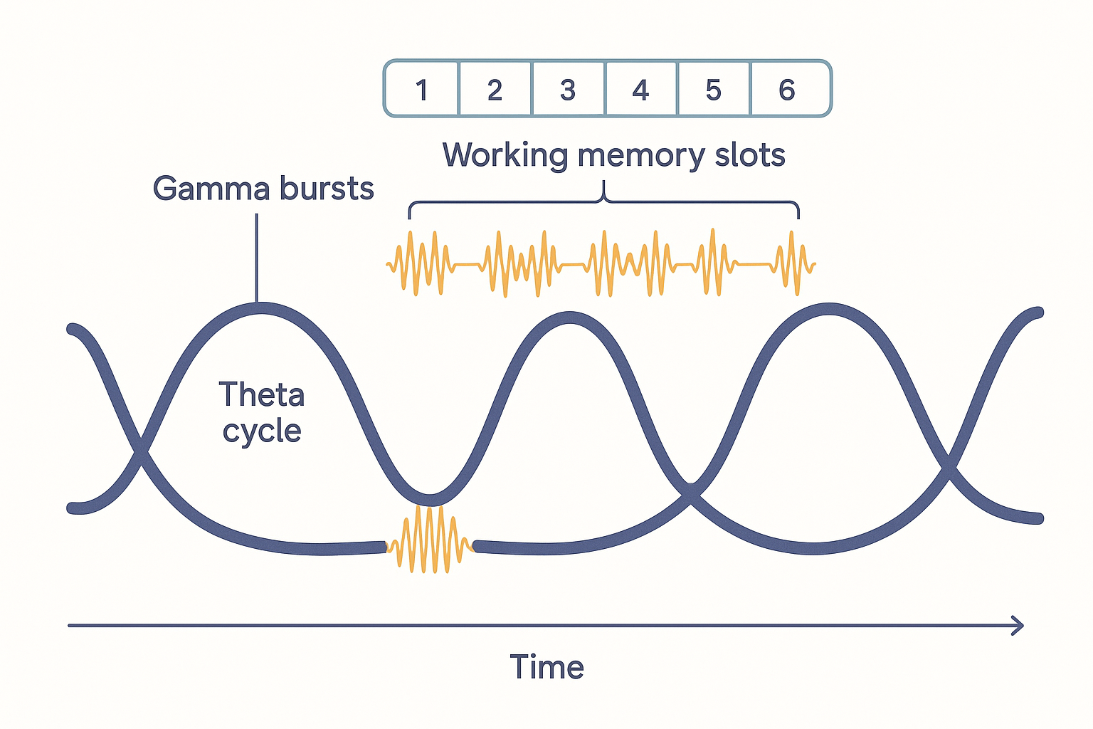 Theta–gamma cross-frequency coupling. The slow theta wave supplies a repeating frame; bursts of fast gamma ride on each cycle, and successive gamma cycles act as ordered "slots" — a candidate physical basis for holding a short sequence of items in working memory.
Taken together — coherence routing the channels, phase coding stamping the timing, cross-frequency coupling nesting fast inside slow — these mechanisms recast oscillations from a passive readout into the brain's temporal operating system: the means by which a fixed anatomical wiring diagram is dynamically reconfigured, millisecond by millisecond, into the specific computation the moment requires.
How brain waves are measured
EEG — cheap, fast, and blurry
The electroencephalogram (EEG) places metal electrodes on the scalp and measures the voltage between them, sampling hundreds to thousands of times a second. Its great strength is temporal resolution: it sees events on the millisecond timescale at which the brain actually operates, which is exactly the timescale of oscillations. Its great weakness is spatial resolution. The signal must pass through the cerebrospinal fluid, skull, and scalp, which smear it badly, so a scalp electrode reports the blurred sum of activity from a broad patch of cortex beneath it. Worse, inferring which deep sources produced a given scalp pattern is the inverse problem — mathematically, infinitely many internal source configurations can produce the same surface map, so EEG can never uniquely localise a generator. EEG tells you when, precisely, and where, only roughly.
Recall the constraints from the opening sections, because they bound what EEG can ever see:
- It reads summed synchronous postsynaptic potentials, dominated by the aligned pyramidal cells of the cortical surface. It is blind to single spikes and largely blind to deep structures (hippocampus, thalamus) whose geometry does not produce a strong open field at the scalp.
- It sees only synchronous populations. A region computing hard but asynchronously contributes little, even though it is intensely active — the metabolically busiest cortex can be electrically quiet.
- It is exquisitely sensitive to artefacts that dwarf the brain signal: eye blinks, muscle tension, heartbeat, and electrical mains hum are all far larger than the microvolt-scale EEG and must be filtered out — a perennial source of bad "brain-wave" claims built on muscle noise.
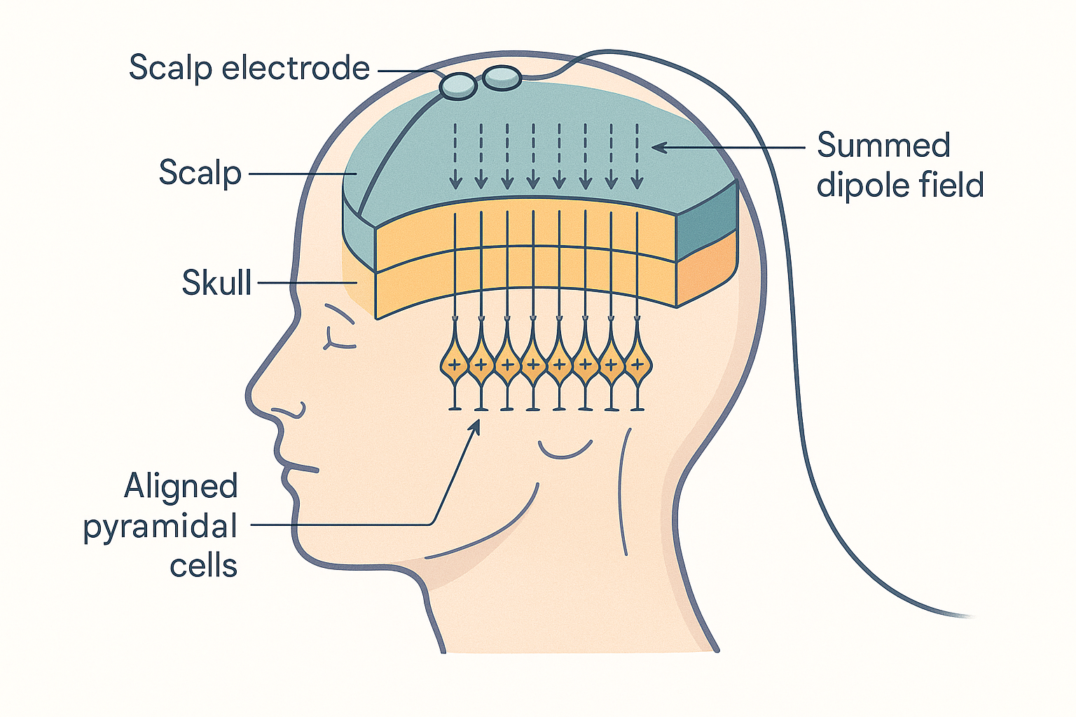 Why the scalp can read the brain: aligned cortical pyramidal cells act as parallel dipoles. Only when millions sum in synchrony does a microvolt-scale field survive the blurring journey through skull and scalp to reach an electrode — and the inverse path back to the source is fundamentally ambiguous.
MEG and the others
Magnetoencephalography (MEG) measures the tiny magnetic fields produced by the same neural currents, using superconducting sensors. Magnetic fields pass through the skull undistorted, so MEG offers better spatial localisation than EEG, and it is especially good at sources tangential to the surface (tucked in the folds, the sulci) where EEG is weakest. But it is enormous, fabulously expensive, and must be run inside a magnetically shielded room — a research tool, not a wearable. Intracranial recording (electrodes placed directly on or in the brain, in epilepsy patients) gives the cleanest, most local oscillations of all and is the source of much of what we know about human gamma and theta–gamma coupling, but it requires surgery.
Event-related potentials — averaging out the rhythm
A related technique deserves a line because it is constantly confused with brain-wave analysis. An event-related potential (ERP) is obtained by presenting the same stimulus many times and averaging the EEG locked to each presentation. Averaging cancels the ongoing background rhythms (which are not time-locked to the stimulus) and leaves the brain's stereotyped response to the event — a sequence of characteristic peaks (named by polarity and timing, e.g. the P300, a positive deflection ~300 ms after a surprising stimulus). ERPs measure the brain's reaction to events; oscillation analysis measures its ongoing rhythms. They are two different lenses on the same EEG.
States and applications: from sleep to neurofeedback
Sleep: the canonical brain-wave staircase
The clearest, best-established use of brain waves is staging sleep, the very thing Dement and Kleitman pioneered. As you fall asleep the EEG descends a staircase of rhythms, each generated by the thalamocortical loop at a deeper level of hyperpolarisation:
- Wake (eyes closed): alpha dominates, with beta when alert.
- N1 (drowsy): alpha fades; slow theta appears. The threshold of sleep.
- N2 (light sleep): sleep spindles and K-complexes punctuate a theta background. The bulk of the night.
- N3 (deep / slow-wave sleep): delta slow waves take over — the restorative depths.
- REM (dreaming): the EEG paradoxically speeds up to look almost awake — low-amplitude, fast, theta-rich — while the body is paralysed and the eyes dart. This is why REM is called "paradoxical sleep."
The functional pay-off is memory consolidation, and it is one of the better-evidenced stories in the field. During slow-wave sleep, the hippocampus replays the day's experiences and, through the coordinated interplay of delta slow waves, thalamic spindles, and hippocampal sharp-wave ripples, transfers them into long-term cortical storage. Spindle density and slow-wave amount predict how much you remember the next day. Brain rhythms are not just a label for sleep stages; they appear to be the machinery by which sleep does its work — the substance of the sleep architecture page.
Neurofeedback: training your own rhythms
Neurofeedback measures your EEG in real time and feeds back a signal — a tone, a game, a bar on a screen — that rewards you when a target band rises or falls, the idea being that you can learn to self-regulate your own rhythms (boost SMR for calm focus, reduce excess theta in ADHD, raise alpha for relaxation).
Calibration — neurofeedback
The premise is biologically real (the brain can learn to modulate its rhythms through feedback), and the technique is benign. But the clinical evidence is mixed and heavily confounded. The most rigorous trials — especially in ADHD, its flagship application — repeatedly find that neurofeedback beats doing nothing but fails to beat a convincing sham (fake feedback) in well-blinded designs, implying much of the benefit is expectation, attention, and the structured practice of sitting still and concentrating, rather than the specific rhythm being trained. It is plausibly a useful relaxation-and-attention practice; it is not the precise "rewire your brain wave by wave" tool it is often sold as.
Binaural beats: an honest look
Play a 200 Hz tone in one ear and a 210 Hz tone in the other, and your brain perceives a phantom 10 Hz "beat" — the binaural beat, a frequency difference computed in the auditory brainstem. The marketing claim is entrainment: that listening to a binaural beat at, say, 10 Hz will pull your whole brain into a 10 Hz alpha state ("relaxation"), or 5 Hz theta ("meditation"), or 40 Hz gamma ("focus").
Calibration — binaural beats
The perceptual phenomenon is real, but the strong claim — that the beat entrains cortical oscillations and thereby reliably changes your mental state — is weakly and inconsistently supported. Studies measuring EEG during binaural beats often find little or no genuine entrainment of cortical rhythms to the beat frequency, and reported effects on mood, anxiety, or attention are small, inconsistent across studies, and hard to separate from simple relaxation, expectation, and the calming effect of listening to gentle sound through headphones. There is no good evidence that the specific beat frequency does what the labels claim. Contrast this with direct sensory entrainment below, where an actual flickering or pulsing stimulus does measurably drive cortical rhythms — the binaural trick is far more indirect and far weaker.
Meditation and the alpha/theta story
Experienced meditators reliably show increased alpha and theta power during practice, and long-term practitioners of certain styles show striking, sustained high-amplitude gamma synchrony — among the strongest gamma ever recorded in healthy humans, in the famous studies of Tibetan monks.
Calibration — meditation
The EEG correlates of meditation are robust and replicated: practice genuinely shifts the spectrum (more alpha/theta during relaxed focused attention; trained gamma in advanced practitioners). What is not established is the causal arrow implied by "meditate to produce theta and theta produces benefits." The rhythm changes are most parsimoniously read as signatures of the mental state the practice cultivates — relaxed, internally directed, inhibiting external distractors (exactly what an alpha gate would do) — rather than the cause of the benefits. The waves are the readout of a skill, not a shortcut around acquiring it.
Gamma entrainment (40 Hz): the most interesting current research
The single most exciting brain-wave story right now is sensory gamma entrainment in Alzheimer's disease. Unlike binaural beats, this uses a real, rhythmic sensory drive — light flickering at exactly 40 Hz and/or sound clicking at 40 Hz — which genuinely entrains cortical gamma (the brain follows the flicker, a phenomenon long known as the steady-state response). The pioneering work (Li-Huei Tsai's group at MIT, branded GENUS — gamma entrainment using sensory stimulation) found, first in mice, that 40 Hz stimulation reduced amyloid-beta plaques, engaged the brain's immune cells (microglia) and waste-clearance pathways, and improved cognition — a remarkable result given that gamma-generating PV interneurons are among the first cells to fail in Alzheimer's.
Calibration — 40 Hz gamma
This is a promising, biologically grounded line of research — not yet a proven therapy. The mouse data are strong and mechanistically rich; the human data are early. Small phase-I/II trials report that daily 40 Hz light-and-sound is safe and well-tolerated, with preliminary signals of slowed brain atrophy, preserved functional connectivity, and modest cognitive and sleep benefits versus controls — but these are small, partly unblinded studies, and at least one independent attempt has struggled to reproduce the headline amyloid-clearance effect. A pivotal phase-III trial (Cognito Therapeutics) is underway and will be the real test. Treat it as the most credible brain-wave intervention on the horizon and the one worth watching — while remembering that "in phase-III trials" is not the same as "proven to work." It is the clearest example on this page of an oscillation being used not as a readout but as a lever on the tissue.
Putting it all together
- A neural oscillation is summed, synchronised postsynaptic potentials — not spikes. EEG is visible only because the cortex's pyramidal cells are aligned like an orchard and, when they idle in unison, their tiny dipoles add into a field readable at the scalp. Big slow waves mean synchronous idling; small fast waves mean busy, independent work.
- Rhythms are generated by inhibition. A local excitation–inhibition loop, paced by GABAergic PV fast-spiking interneurons (PING/ING), sets the fast bands; the larger thalamocortical loop, retuned by brainstem arousal chemistry, sets alpha and the descending sleep rhythms via the thalamic reticular nucleus and rebound bursting. The brain's clocks are made of brakes.
- The five bands map to states and functions: delta (deep-sleep restoration and memory consolidation), theta (hippocampal memory and navigation, REM), alpha (relaxed-wakefulness inhibitory gating — suppressing the irrelevant), beta (alert engagement and motor status-quo), gamma (active perception, attention, and feature binding, driven by PV interneurons). Slower = bigger and more global; faster = smaller and more local.
- Rhythms do real computational work: communication-through-coherence routes information by phase-aligning regions; phase coding stamps meaning onto spike timing; theta–gamma cross-frequency coupling nests fast slots inside a slow frame — a candidate basis for the ~7-item limit of working memory.
- Measurement is a trade-off: EEG gives millisecond timing but blurry, surface-biased, synchrony-only, artefact-prone localisation (the inverse problem); MEG and intracranial recording sharpen the picture at great cost; ERPs average the rhythm away to isolate the response to events.
- Calibrate the applications honestly: sleep staging and memory-consolidation rhythms are solid; neurofeedback and binaural beats are weak-to-mixed once properly controlled; meditation's wave changes are real signatures but not proven causes; and 40 Hz sensory gamma entrainment is the one genuinely promising intervention — mechanistically deep, early in humans, and now in phase-III trials.
The unifying idea: the cognition page gave you the brain's wiring and chemistry — the spatial machine. This page gives you its timing — the temporal machine layered on top. Neurons do not merely connect; they coordinate in time, and that coordination — who fires together, at what rhythm, in what phase — is how a fixed anatomy becomes a flexible, moment-to-moment computer. Brain waves are not noise the brain happens to make. They may be the medium in which the brain thinks.
Related Compounds & Deep Dives
Set the rhythm (GABA / inhibition — the pacemakers of fast oscillations)
- GABA & E/I balance — the inhibitory system that paces every fast rhythm; PV interneurons are the brain's metronomes.
- Phenibut — GABA-B agonist; like alcohol and benzodiazepines, GABAergic drugs slow and reshape the EEG toward higher-amplitude slow activity.
- Apigenin — flavonoid with benzodiazepine-site (GABA-A) activity; mild sedative, EEG-slowing direction.
- CBD — modulates cortical excitability and the E/I balance that underlies oscillation.
Alpha and relaxed wakefulness
- L-theanine — the best-evidenced "alpha enhancer": reliably increases occipital alpha power, the rhythm of relaxed, alert, eyes-closed wakefulness.
- Magnesium — tunes NMDA signal-to-noise (see the deep dive); supports the E/I balance that clean oscillations depend on.
Sleep rhythms (delta, spindles, slow waves)
- Melatonin — shifts state toward sleep, indirectly favouring the thalamocortical descent into spindles and delta.
- Glycine — inhibitory transmitter that lowers body temperature and improves slow-wave sleep quality.
Gamma, attention, and the PV/cholinergic side
- Acetylcholine / choline / citicoline — cholinergic tone shifts the thalamocortical loop from sleep rhythms toward fast, desynchronised, gamma-friendly waking activity.
- Nicotine — nicotinic activation of interneurons modulates gamma and attention.
- Caffeine — adenosine antagonist; suppresses the slow waves of drowsiness and pushes the EEG toward alert beta (see the cognition page).
- Creatine — buffers the brain's huge energy cost of sustaining fast oscillations under load (see cellular energy).
Rhythm disruptors / psychedelics
- Psilocybin — desynchronises normal cortical rhythms (notably reducing alpha), a candidate mechanism for the dissolution of ordinary perceptual binding.
Related foundations
- Neuroscience of Cognition — the spatial wiring and chemistry this page adds timing to.
- GABA & E/I balance — the inhibitory interneurons that generate the rhythms.
- Sleep Architecture — the brain-wave staircase of the night and memory consolidation, in full.
- Autonomic Nervous System — the brainstem arousal systems that retune the thalamocortical loop between waking and sleep.
- Cellular Energy — why sustaining fast oscillation is metabolically expensive, and where adenosine (the tiredness signal) comes from.