The Thymus
The strangest organ in the body: a gland that does its most important work before you are grown, then deliberately dismantles itself. This page explains what the thymus actually is — the "schoolhouse" where every T-cell is trained to tell self from non-self — why it shrinks and turns to fat as you age, why that shrinkage may be one of the deepest clocks of immune ageing, and why a small, hyped family of peptides claims to wind it back.
The thymus spent most of medical history as a puzzle. The Greeks named it after thymos — spirit, the seat of emotion — and for two millennia nobody knew what it did. Anatomists noticed it was large in children and nearly gone in adults, and concluded it was a vestigial organ: a leftover with no real function, sometimes blamed (wrongly) for cot death. As late as the 1950s, surgeons irradiated children's thymuses as if they were harmless.
That ended in 1961, when the Australian-British immunologist Jacques Miller, working in London, removed the thymus from newborn mice and watched their immune systems collapse — they could not reject skin grafts, made few lymphocytes, and died of infection. Miller had discovered that the thymus is where one entire arm of the immune system is made: it is the organ that produces and educates T-cells (the "T" stands for thymus). It was the last organ in the human body to have its function discovered. The next four decades filled in how the education works — the twin selection steps, and, around the 1990s–2000s, the discovery of the gene AIRE and the mechanism of central tolerance (how the body learns not to attack itself). Today the thymus is a live frontier of longevity science: researchers are trying to regrow it (the small TRIIM trial reported apparent regeneration and even epigenetic age reversal), to build thymic organoids in the lab, and — at the speculative fringe — to nudge it chemically with peptide "bioregulators" like Vilon. The vestigial organ turned out to be one of the most important and most under-appreciated glands you own.
Why this page exists
The immune system page introduces the cells of defence; the lymphatic system page maps the vessels and nodes they travel through. This page zooms in on a single organ that sits upstream of both: the factory and training academy for T-cells, the immune system's precision-targeting arm. It earns its own page for three reasons.
First, the thymus is the clearest example in the whole body of a profound biological idea: the immune system is not born knowing what to attack — it has to be taught, cell by cell, not to attack you. That teaching happens almost entirely inside the thymus, and the logic of it (build random weapons, then ruthlessly destroy the ones that point inward) is one of the most elegant and brutal processes in physiology.
Second, the thymus is the body's most dramatic example of programmed ageing. Unlike most organs, which decline gradually and reluctantly, the thymus begins shrinking in childhood and is mostly replaced by fat by middle age — on purpose. Understanding why is central to understanding immunosenescence (the ageing of the immune system) and inflammaging, themes that run through the inflammation page and the entire longevity discussion.
Third, it is the organ at the centre of one of the database's most-discussed peptide families — the Khavinson bioregulators, Vilon and Thymalin. You cannot judge the claim that a molecule "re-engages the thymus" without first knowing precisely what the thymus does. We build that picture from the anatomy up.
Where it sits: a primary lymphoid organ
Before the anatomy, place the thymus in the hierarchy. Immune (lymphoid) organs come in two tiers, and the distinction is the key to the whole page:
- Primary (central) lymphoid organs are where immune cells are born and trained. There are exactly two: the bone marrow (which makes all blood cells, and where B-cells mature) and the thymus (where T-cells mature). These are schools.
- Secondary (peripheral) lymphoid organs are where mature immune cells do their job — meeting pathogens and launching responses. These are the lymph nodes, the spleen, the tonsils, and the gut's lymphoid patches. These are the battlefields. (They are the subject of the lymphatic system page.)
flowchart TD
    BM["BONE MARROW<br/>(makes all blood cells)"] -->|"B-cell progenitors mature here"| BCELL["Mature naive B-cells"]
    BM -->|"T-cell progenitors leave<br/>BEFORE maturing"| THY["THYMUS<br/>(T-cell training school)"]
    THY -->|"educated, self-tolerant"| TCELL["Mature naive T-cells<br/>(CD4+ and CD8+)"]
    BCELL --> SEC["SECONDARY ORGANS<br/>lymph nodes · spleen · tonsils · gut patches"]
    TCELL --> SEC
    SEC --> FIGHT["Meet antigen → mount<br/>the actual immune response"]
    subgraph PRIMARY["PRIMARY (central) — where cells are MADE + TRAINED"]
      BM
      THY
    end
The thymus's defining oddity is here: T-cell precursors are made in the bone marrow but are not trained there. They are shipped, immature and dangerous, to a dedicated organ — the thymus — purely to be educated. No other cell lineage gets its own training organ. That tells you how dangerous a badly-trained T-cell is, and how much machinery the body invests to prevent one.
Anatomy and histology: the building
The thymus sits in the anterior mediastinum — the central compartment of the chest, directly behind the sternum (breastbone) and just in front of and above the heart and great vessels. In a child it is a soft, pinkish-grey, two-lobed organ draped over the top of the heart. It reaches its greatest absolute size — around 30–40 g — at puberty, then begins its long retreat (below).
The internal architecture is the part that matters, because structure is function here. Each lobe is divided into thousands of lobules, and every lobule has the same two-zone layout — an outer cortex and an inner medulla — and a maturing T-cell physically travels from one to the other as it is educated.
- The cortex is the dense, dark-staining outer zone, packed tightly with immature T-cells (called thymocytes while inside the thymus). This is where the first exam happens.
- The medulla is the paler, less crowded inner zone, where the second, more stringent exam happens, and from which the survivors exit into the blood.
The walls and scaffolding of this building are not made of ordinary connective tissue but of a remarkable cell type:
- Thymic epithelial cells (TECs) — the "teachers." They form a three-dimensional sponge-like meshwork that the thymocytes crawl through, and they are physically different in the two zones:
- Cortical TECs (cTECs) run the cortex and conduct positive selection (test 1).
- Medullary TECs (mTECs) run the medulla and conduct negative selection (test 2). They have a near-magical property we return to: they display fragments of the entire body inside the thymus.
- Dendritic cells and macrophages are also present — dendritic cells help test thymocytes against self, and macrophages eat the enormous number of thymocytes that fail (almost all of them).
- Hassall's corpuscles are distinctive onion-like whorls of flattened, keratinised mTECs found only in the medulla. Long a histological curiosity (they are the textbook landmark for identifying thymic tissue under the microscope), they are now thought to help instruct the production of regulatory T-cells (below).
- The blood–thymus barrier wraps the cortical blood vessels in a tight epithelial sheath, so that circulating foreign antigens are kept out of the cortex. This is deliberate: during positive selection, thymocytes must be tested against self antigens only, with no confusing foreign material present. The barrier keeps the first exam "clean."
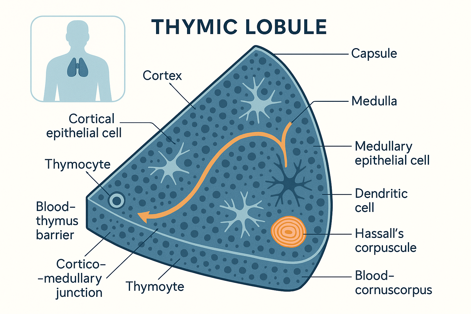 The thymic lobule: thymocytes enter at the cortico-medullary junction, are tested in the dense cortex (positive selection by cortical TECs), then migrate inward to the medulla (negative selection by medullary TECs and dendritic cells) before the survivors exit to the blood.
The core function: the T-cell schoolhouse
Now the central process — and to follow it you need three concepts defined first.
A T-cell is a white blood cell that, unlike the antibody-making B-cell, works by direct inspection: it physically touches other cells and reads what they are displaying on their surface, then either kills them or issues commands. It is the immune system's precision instrument.
The T-cell receptor (TCR) is the unique sensor on each T-cell's surface — its "key." Crucially, each T-cell builds its TCR by randomly shuffling and splicing gene segments (a process called V(D)J recombination). The randomness is the entire point: by generating millions of different TCRs, the body ensures that some T-cell, somewhere, will recognise any pathogen it ever meets, including ones that don't yet exist. But randomness has a catastrophic side effect — a randomly built receptor is just as likely to recognise your own tissues as a virus. That is the problem the thymus exists to solve.
MHC (major histocompatibility complex) molecules are the "display trays" present on the surface of your cells. A cell constantly chops up samples of the proteins it contains and presents the fragments on MHC molecules for T-cells to inspect — like a shop displaying samples of everything in the back room. There are two kinds: MHC class I (on almost every cell, displaying what's inside — so a virus-infected cell reveals viral fragments) and MHC class II (on specialised immune cells, displaying what they've eaten). A T-cell never sees free-floating antigen; it only ever reads antigen presented on an MHC tray. This is the deep rule the thymus must enforce.
So a usable T-cell must pass two tests, which are genuinely opposite in spirit:
- It must be able to read the MHC trays at all — a TCR that can't engage your own MHC molecules is useless, because MHC is the only language T-cells speak. This is positive selection, and it establishes MHC restriction (the T-cell is locked to recognising antigen only when shown on self-MHC).
- It must not react to your own body — a TCR that binds too strongly to your own self-antigens (presented on those trays) is a loaded autoimmune weapon and must be destroyed. This is negative selection, and it establishes central tolerance (the T-cell will not attack self).
The thymus runs both, in sequence, in its two zones.
Test 1 — positive selection (the cortex): "can you read self-MHC?"
A thymocyte enters at the junction between cortex and medulla, double-negative (it carries neither of the two co-receptors, CD4 or CD8, yet), rearranges its TCR genes, and migrates outward into the cortex now expressing both co-receptors (double-positive). There, cortical TECs present it with self-MHC molecules carrying self-peptides. The test is simple and binary:
- If the thymocyte's randomly-built TCR can bind self-MHC with at least a weak, adequate affinity → it receives a survival signal. It can "read the language." It survives, and commits to one lineage: bind MHC class I → becomes a CD8+ ("cytotoxic," cell-killing) T-cell; bind MHC class II → becomes a CD4+ ("helper," command-issuing) T-cell.
- If the TCR cannot engage self-MHC at all → it gets no survival signal and dies quietly by neglect (a fate called death by neglect). A receptor that can't read the trays is worthless, so it is discarded.
The great majority of thymocytes fail here — most randomly-generated receptors simply don't fit self-MHC. This is the first cull.
Test 2 — negative selection (the medulla): "do you attack self?"
The survivors migrate inward to the medulla for the harder, more dangerous exam. Here the question reverses: not "can you bind self?" but "do you bind self too strongly?" Medullary TECs and dendritic cells present a vast library of self-antigens, and the rule is:
- If the thymocyte binds a self-antigen too strongly → it is a potential autoimmune attacker. It is ordered to die (by apoptosis, programmed cell death) and is eaten by macrophages. This deletion of self-reactive clones is central tolerance.
- If it binds self only weakly or not at all → it passes, and is cleared for export.
But this raises an obvious problem, and its solution is one of immunology's most beautiful discoveries. How can the medulla test a thymocyte against, say, insulin (made only in the pancreas) or thyroglobulin (made only in the thyroid)? Those proteins aren't in the thymus. If they're never shown, T-cells that attack the pancreas or thyroid would slip through.
The answer is AIRE. The autoimmune regulator (AIRE) is a transcription factor — a master gene-switch — expressed by medullary TECs, and it does something extraordinary: it forces those cells to manufacture small amounts of proteins from all over the body — insulin, thyroglobulin, retinal proteins, muscle proteins — genes that have no business being switched on in a chest gland. This is called promiscuous gene expression. In effect, AIRE makes the medulla display a miniature catalogue of the entire self, so that every developing T-cell can be tested against tissues it will never otherwise encounter until it's too late. A thymocyte that would have attacked the pancreas meets pancreatic insulin here, in the safety of the thymus, and is deleted before it ever leaves.
The clinical proof is stark: people born with a broken AIRE gene develop APECED (also called APS-1), a devastating multi-organ autoimmune disease — their immune systems attack the parathyroids, adrenals, and other tissues precisely because those self-antigens were never shown in the thymus. One faulty gene-switch, and central tolerance fails.
The output, and the brutal arithmetic
A small fraction of thymocytes — those that can read self-MHC (passed test 1) but do not over-react to self (passed test 2) — graduate. They become single-positive naïve T-cells (either CD4+ or CD8+, "naïve" meaning they have never yet met their target antigen), and they exit via the medullary blood vessels into circulation, heading for the lymph nodes and spleen to wait for their pathogen.
A special subset deserves mention: some CD4+ thymocytes with intermediate self-reactivity are not deleted but converted into regulatory T-cells (Tregs) — identifiable by the master gene FOXP3 — which actively suppress immune responses and police tolerance out in the body (Hassall's corpuscles are implicated in instructing their production). The thymus thus exports both the soldiers and the military police.
Now the statistic that captures the whole process: of all the thymocytes that enter and attempt this gauntlet, only about 2–5% survive. Roughly 95–98% are destroyed — the vast majority for failing positive selection, plus all the dangerously self-reactive ones removed by negative selection. The thymus is, by design, one of the most lethal places in the body — a school that expels almost every pupil, because the cost of graduating even one self-reactive T-cell (autoimmune disease) is higher than the cost of throwing away millions of usable ones. It is profligate on purpose.
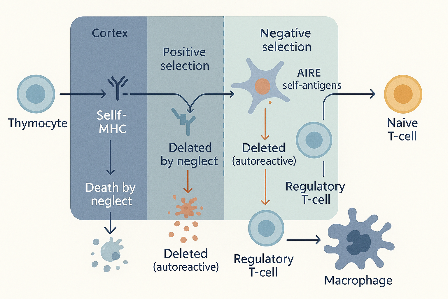 The selection gauntlet: positive selection keeps T-cells that can read self-MHC; negative selection deletes those that attack self (with AIRE displaying a catalogue of whole-body antigens). Only ~2–5% survive both tests to become safe, naive T-cells.
The endocrine side: thymic hormones and their cofactors
The thymus is usually filed as a lymphoid organ, but it is also, quietly, an endocrine one — it secretes its own hormones (peptide signals released into the blood to act elsewhere), which is the bridge between this page and the hormones page. The thymic epithelial cells don't just train T-cells by contact; they release soluble factors that tune T-cell maturation and signal the wider immune and endocrine systems. The main ones:
- Thymulin — a nonapeptide (nine-amino-acid hormone) and the most cofactor-relevant of all, because thymulin is biologically inactive without zinc. The peptide must bind a single zinc ion to fold into its active shape; the zinc-free form does nothing. This makes thymulin a direct molecular reason why zinc deficiency cripples immunity — low zinc means low active thymulin, even if the peptide itself is present. It is one of the cleanest examples in the body of a trace mineral acting as an obligatory cofactor for a hormone, and the mechanistic basis for zinc's reputation as an immune nutrient. Active thymulin promotes T-cell differentiation and helps set immune tone.
- Thymosin alpha-1 — a peptide that boosts T-cell maturation and activation; it is the one thymic peptide that became an actual licensed drug (used in some countries as an immune stimulant in hepatitis and as an adjuvant).
- Thymopoietin — influences thymocyte differentiation (and, separately, neuromuscular transmission).
- Thymic humoral factor and others — additional peptides supporting peripheral T-cell function.
The endocrine traffic runs both ways — the thymus is exquisitely sensitive to hormones from elsewhere, which is the key to understanding why it shrinks. The major external controls (each a cross-link to the hormones and metabolism pages):
- Cortisol / glucocorticoids (the stress link). Thymocytes are extraordinarily sensitive to glucocorticoids — cortisol triggers their apoptosis. This is why acute stress, illness, or steroid treatment causes rapid, visible thymic shrinkage ("stress involution" or "accidental involution"); the thymus can lose much of its mass within days of a severe stressor and regrow afterward. The HPA stress axis has a direct dial on the size of your immune training organ.
- Sex steroids (the puberty link). Androgens and oestrogens drive thymic involution — which is why the great shrinkage accelerates at puberty, exactly when sex steroids surge. The proof is dramatic and reversible: castration (androgen removal) regrows the thymus in animals and men undergoing androgen-deprivation therapy, and blocking sex steroids is an active experimental strategy for thymic regeneration.
- Growth hormone and IGF-1 (the growth link). These support thymic size and naïve T-cell output, which is the rationale behind the TRIIM regeneration trial below.
- Leptin (the nutrition link). Leptin, the fat-derived "I am fed" hormone from the hormones page, supports the thymus — which is why starvation and severe malnutrition shrink it (leptin falls) and immunity collapses. The thymus is a luxury organ the body trims first when energy is scarce.
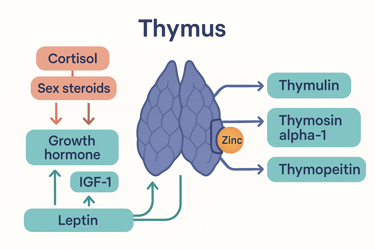 The thymus is also an endocrine gland: it secretes thymulin (active only when bound to zinc), thymosin alpha-1 and thymopoietin, while itself being shrunk by cortisol and sex steroids and supported by growth hormone, IGF-1 and leptin.
Involution: the organ that dismantles itself
Here is what makes the thymus unique among organs: it ages first, and on purpose. Most organs decline slowly and grudgingly over a lifetime. The thymus does the opposite — it reaches peak function early and then deliberately disassembles, a programmed process called thymic involution.
The timeline, with real numbers:
- The thymus is at its largest relative to body size at birth and in early childhood — proportionally it is never bigger than in a newborn, because building the entire T-cell repertoire from scratch is the immune system's most urgent early task.
- It reaches peak absolute mass (~30–40 g) around puberty.
- From puberty it involutes at roughly 3% per year through early adulthood, slowing to about 1% per year later. The functional, T-cell-producing tissue (the epithelial cortex and medulla) is progressively replaced by adipose (fat) tissue — on an MRI or at autopsy, a middle-aged thymus looks like a pad of fat with islands of shrinking immune tissue.
- By around age 50, the majority of functional thymic tissue is gone; by old age only small active remnants persist amid the fat.
The consequence is a steady fall in naïve T-cell output. (This can be measured: newly-made T-cells carry tiny circular DNA by-products of TCR rearrangement called TRECs — T-cell receptor excision circles — and TREC levels in the blood fall steeply with age, a direct readout of thymic activity.) As the supply of fresh naïve T-cells dries up, the immune system is forced to rely on the fixed pool of memory T-cells it accumulated earlier in life, topped up by sluggish division of existing cells rather than new thymic graduates. This is the heart of immunosenescence — the ageing of the immune system:
- A shrinking TCR repertoire. Fewer new naïve cells means fewer novel receptor specificities. The diversity of the T-cell repertoire — the breadth of threats the immune system can recognise — narrows with age. New pathogens (and new vaccines) find fewer matching T-cells waiting.
- Accumulation of memory and "exhausted" cells. The repertoire fills with memory cells from past infections (chronic viruses like cytomegalovirus consume a startling fraction of an old person's T-cell space) and with senescent, poorly-functioning, "exhausted" cells. The CD4:CD8 ratio often inverts.
- The downstream consequences are exactly the clinical face of immune ageing:
- Poorer vaccine responses — older immune systems struggle to mount fresh responses to new antigens, which is why vaccines are less effective in the elderly and why high-dose formulations exist.
- Greater infection susceptibility and severity — fewer naïve T-cells to respond to novel pathogens (vividly illustrated by the age gradient of COVID-19 mortality).
- Weaker tumour surveillance — T-cells patrol for cancerous cells displaying abnormal antigens on MHC; a depleted, narrowed repertoire watches less well, part of why cancer incidence climbs with age.
- "Inflammaging" — the chronic, low-grade, sterile inflammation of older age (the inflammation page's smouldering fire). A failing thymus contributes: loss of regulatory T-cells and a dysregulated, memory-skewed compartment tilt the system toward background inflammation.
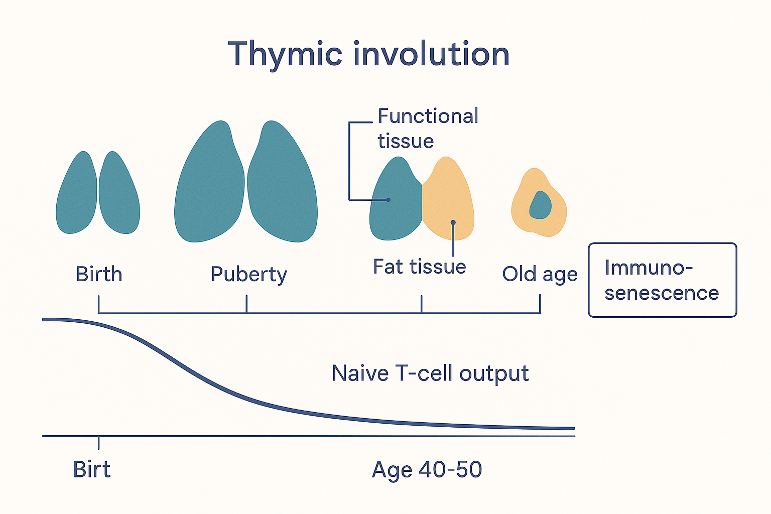 Thymic involution: the thymus peaks at puberty then is steadily replaced by fat, naive T-cell output falls, and the immune system shifts to a fixed, ageing pool of memory cells — the engine of immunosenescence and inflammaging.
Why would evolution build an organ that destroys itself? The leading view is that involution is a trade-off, not a defect. Building the core T-cell repertoire is an early-life job; once a broad library of naïve cells and a stock of memory cells exist, running a large, metabolically expensive, and intrinsically dangerous organ (remember it spends its life generating self-reactive cells that must be culled) yields diminishing returns. The body reallocates the resources. This was a fine bargain when human lifespans were short. In an era of long lifespans, the early shutdown of the thymus leaves us with decades of life on a depleted, ageing immune system — which is precisely why the thymus has become a target for longevity science.
The longevity angle: can the thymus be rebuilt?
If thymic involution is a central clock of immune ageing, the obvious question is whether it can be slowed or reversed. A healthier thymus, the argument goes, would mean more naïve T-cells, a broader repertoire, better responses to new infections and vaccines, sharper tumour surveillance, and less inflammaging — a meaningful lever on healthspan. This is a legitimate, mainstream research goal, and a few honest data points exist (calibrated carefully, because this is an area thick with hype):
- The TRIIM trial (2019). A small study (the Thymus Regeneration, Immunorestoration and Insulin Mitigation trial) gave nine healthy middle-aged men a one-year cocktail of growth hormone (to stimulate the thymus) plus DHEA and metformin (to offset GH's tendency to raise blood sugar). MRI suggested apparent regrowth of functional thymic tissue (fat being replaced by active tissue — the reverse of involution), alongside improved immune markers and, strikingly, an apparent reversal of epigenetic age of about 2.5 years on methylation "clocks." This is genuinely interesting and the first human hint that involution might be reversible. But calibrate hard: it was tiny (n=9), uncontrolled (no placebo group), unblinded, and used growth hormone (which carries its own risks, including cancer-promotion concerns). It is a provocative pilot, not proof. A larger controlled follow-up (TRIIM-X) is the proper test.
- Sex-steroid blockade. Because sex steroids drive involution, blocking them regrows the thymus — demonstrated in men on androgen-deprivation therapy. The effect is real but the intervention (chemical castration) is obviously not a general longevity tool; it mainly proves the principle that involution is not irreversible.
- Thymic organoids and bioengineering. Researchers are growing thymic epithelial tissue in the lab and engineering TECs from stem cells, aiming eventually to rebuild or supplement thymic function — early-stage, but a serious long-term direction.
The bioregulator tie-in (read with calibration)
This brings us to the family of compounds that put the thymus on the biohacking map: the Khavinson peptide bioregulators, specifically Vilon (the dipeptide Lys-Glu) and Thymalin (the crude thymic peptide extract Vilon was distilled from). The claim is that these short thymic peptides "re-engage" the aged thymic epithelium — signalling the involuted, fat-infiltrated gland to resume training T-cells and to shift cytokine output back toward a younger profile.
Everything on this page explains why such a target is plausible in principle: the thymus is genuinely the right organ to address if you want to fight immunosenescence; it is genuinely responsive to peptide and hormonal signals; and its epithelial cells (the cells the peptides are claimed to act on) are genuinely the controllers of the whole process. The target is sound — that part is mainstream immunology, not Khavinson lore.
What this page will not do is endorse the leap from "good target" to "this dipeptide hits it." The evidence that a Lys-Glu peptide actually regrows or re-engages a human thymus is thin — largely single-lab, mostly rodent and cell-culture, with no human trial, no pharmacokinetics, and the most-quoted human/clinical results (the COVID survival figures) belonging to the crude extract Thymalin rather than to Vilon itself. The full, careful evaluation — what stands up, what is misattributed, and where the hype outruns the data — is the subject of the Vilon deep dive; this page deliberately defers the mechanism and evidence argument there. Read it before forming a view. The honest summary for this page: the thymus is exactly the organ you would want to rejuvenate, and that is precisely why it attracts both serious science (TRIIM, organoids) and over-confident peptide marketing.
Putting it all together
- The thymus is a primary lymphoid organ — one of only two (with bone marrow) where immune cells are trained. It sits behind the sternum, above the heart, and exists to educate T-cells (the immune system's precision arm). Long dismissed as vestigial, its function was discovered only in 1961 (Jacques Miller).
- Structure is function: each lobule has an outer cortex (where positive selection happens, run by cortical TECs) and an inner medulla (where negative selection happens, run by medullary TECs and dendritic cells, with Hassall's corpuscles and the blood–thymus barrier as supporting features).
- T-cells build random receptors (TCRs), which makes them able to recognise any pathogen but also dangerously likely to attack self. The thymus solves this with two tests: positive selection (keep only T-cells that can read self-MHC → MHC restriction) and negative selection (delete T-cells that bind self too strongly → central tolerance).
- AIRE lets medullary TECs display a catalogue of whole-body self-antigens inside the thymus, so even tissue-specific proteins (insulin, thyroglobulin) can be used to weed out autoreactive cells; a broken AIRE gene causes multi-organ autoimmunity (APECED). The thymus also exports regulatory T-cells (FOXP3 Tregs).
- The arithmetic is brutal: ~95–98% of thymocytes die during selection — the cost of never graduating a self-reactive cell.
- The thymus is also endocrine: it secretes thymulin (active only when bound to zinc — the clean molecular reason zinc deficiency wrecks immunity), thymosin alpha-1, and thymopoietin. It is in turn controlled by hormones — cortisol and sex steroids shrink it, GH/IGF-1 and leptin support it — which is why stress, puberty, and starvation all reduce thymic size.
- Involution is the headline: the thymus is largest relative to the body at birth, peaks at puberty, then is progressively replaced by fat (~3%/year, then ~1%/year), with most function gone by ~50. Falling naïve T-cell output (measurable as falling TRECs) narrows the TCR repertoire and shifts the immune system onto an ageing pool of memory cells — immunosenescence: worse vaccine responses, more infections, weaker tumour surveillance, and inflammaging.
- Reversing involution is a real longevity target — the small, uncontrolled TRIIM trial hinted at thymic regrowth and epigenetic age reversal with GH+DHEA+metformin; sex-steroid blockade and thymic organoids are other avenues. The Vilon/Thymalin bioregulators claim to re-engage the thymus — a plausible target attached to thin evidence; the deep dive does the careful accounting.
The unifying idea: the thymus is the organ that teaches your immune system not to attack you — and it is the first organ to retire. Almost everything about ageing immunity, from why old people respond poorly to vaccines to why cancer and inflammation climb with age, traces back to a gland that finished its main work in your youth and has been quietly turning to fat ever since. That is what makes it such a compelling — and such a contested — target.
Related Compounds & Deep Dives
The thymic bioregulators (the direct tie-in)
- Vilon — Deep Dive — the Lys-Glu dipeptide pitched as the thymus bioregulator; full evaluation of the claim that it re-engages the aged thymic epithelium.
- Pinealon & Epithalon — Deep Dive — the sister Khavinson peptides (pineal/brain); the same paradigm, mechanism hypothesis, and evidence calibration that frame Vilon.
- Vilon — compound sheet for the thymus peptide.
- Thymalin — the crude thymic extract Vilon was distilled from; the parent of the "doubled COVID survival" claim (extract data, not Vilon data).
Cofactors of thymic function
- Zinc — the obligatory cofactor for thymulin; its zinc-bound form is the active hormone, making zinc directly load-bearing for T-cell maturation.
- Selenium — broader immune and antioxidant cofactor relevant to T-cell function and thymic redox state.
Hormonal controllers
- Growth hormone — supports the thymus and the agent used in the TRIIM regeneration trial.
- Pregnenolone / Progesterone — upstream of the cortisol and sex-steroid signals that drive thymic involution.
Repair-stack neighbours
- BPC-157 — the bioregulator-stack partner most often paired with Vilon (gut and tissue repair from a different angle).
- Melatonin — pineal hormone with immune-supportive effects, linked to the same Khavinson longevity stack via epitalon.
Related foundations
- The Immune System — the T-cells the thymus trains, in the context of the whole defensive network.
- The Lymphatic System — the secondary lymphoid organs (nodes, spleen) where the thymus's graduates do their work.
- Hormones & the Endocrine System — the cortisol, sex-steroid, GH/IGF-1 and leptin signals that control thymic size.
- Inflammation — "inflammaging," the chronic low-grade inflammation that a failing thymus helps drive.